Figure 4.

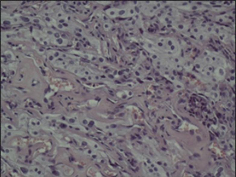
Micrograph of a hematoxylin and eosin-stained tumor specimen (200×) shows moderate cellularity. Meningothelial cells are arranged in small nests and cords. Numerous small blood vessels show marked hyalinization

Micrograph of a hematoxylin and eosin-stained tumor specimen (200×) shows moderate cellularity. Meningothelial cells are arranged in small nests and cords. Numerous small blood vessels show marked hyalinization