Figure 3.

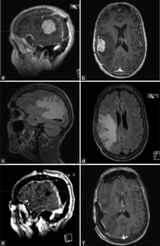
Pre-operative MRI of brain, showing a new enhancing dural based lesion anterior to the prior resection cavity (a-d). Immediate post-operative MRI of brain, demonstrating gross total resection of the lesion (e and f)

Pre-operative MRI of brain, showing a new enhancing dural based lesion anterior to the prior resection cavity (a-d). Immediate post-operative MRI of brain, demonstrating gross total resection of the lesion (e and f)