Figure 3.

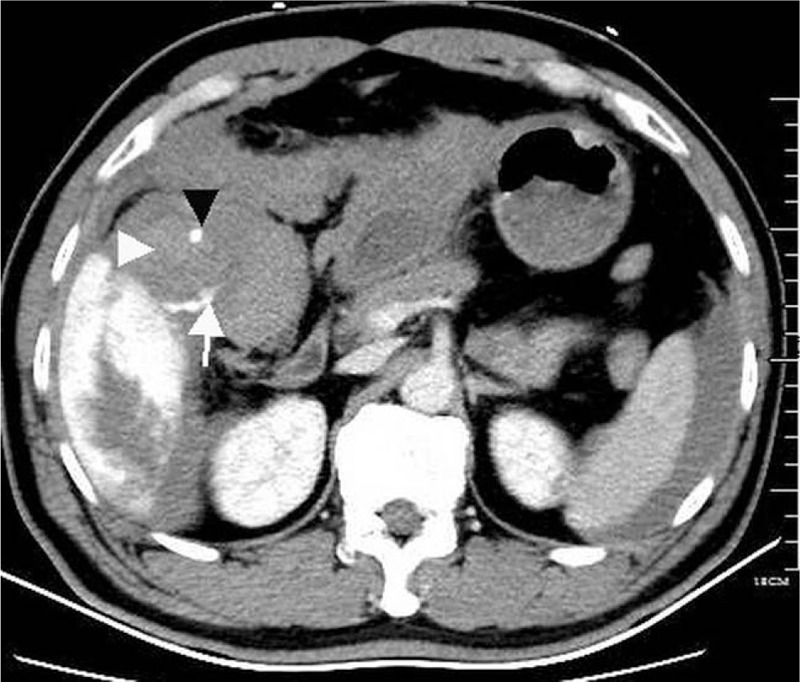
CT shows clots (white arrowhead) around a gallstone (black arrowhead) in the gallbladder and contrast extravasation from the hemangioma to the gallbladder (white arrow). CT = computed tomography.

CT shows clots (white arrowhead) around a gallstone (black arrowhead) in the gallbladder and contrast extravasation from the hemangioma to the gallbladder (white arrow). CT = computed tomography.