Figure 1.

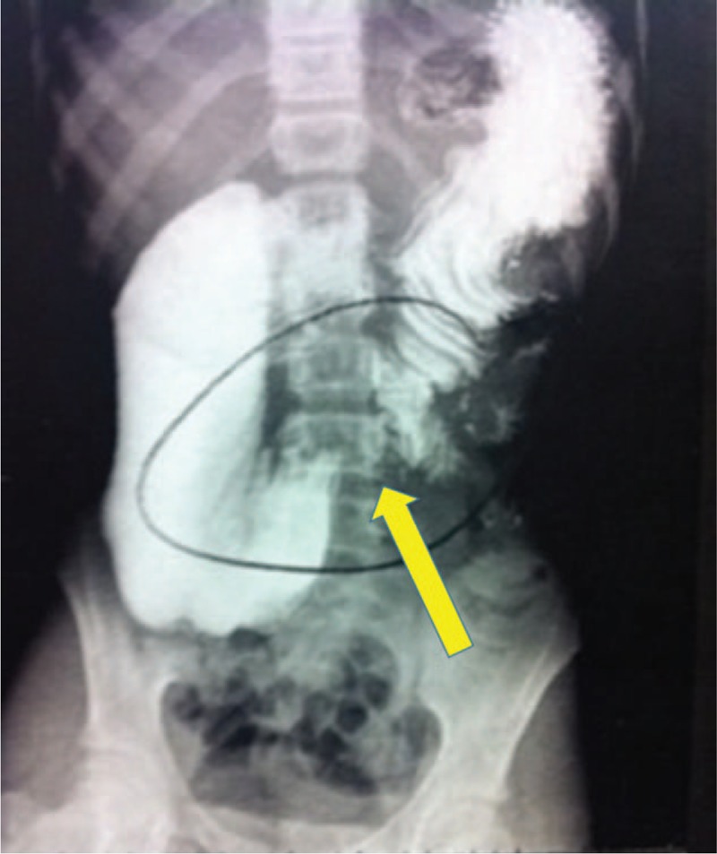
Contrasted abdominal x-ray showing duodenal dilatation with a progression stop of the contrast in the transition between 3rd and 4th duodenal portions.

Contrasted abdominal x-ray showing duodenal dilatation with a progression stop of the contrast in the transition between 3rd and 4th duodenal portions.