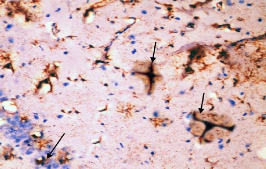
Figure 13.

Immunohistochemical staining for the demonstration of GFAP in the hippocampus of an adult diabetic rat showing many GFAP-positive astrocytes with thick, long and intensely stained processes in the molecular layer and pyramidal layer (arrows). GFAP, ×400
