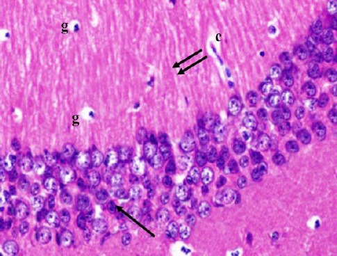
Figure 2.

A photomicrograph of a coronal section of the hippocampus of an adult control rat showing small pyramidal cells of CA1 region (arrow) and molecular layer with the well apparent processes of pyramidal cells (double arrows), glial cells (g) and capillaries (c). H & E; X 400
