
Fig. 8.
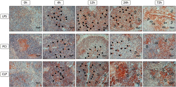
CXCR4 expression in the spleens. At the time point indicated, mice were sacrificed and the spleens were collected for immunohistological analysis. Representative photomicrographs from one of 4–6 different tissue samples stained for CXCR4 expression are shown (red-brown color, counterstaining with hematoxylin; original magnification: 400×). 0 h, 4 h, 12 h, 24 h and 72 h were chosen as representative time points to depict the course. Additionally, arrowheads were added to mark the CXCR4 positive cells that were engulfed by the tingible body macrophages and larger arrows were used to show the infiltrating immune cells exhibiting membrane-bound CXCR4 expression at later time points