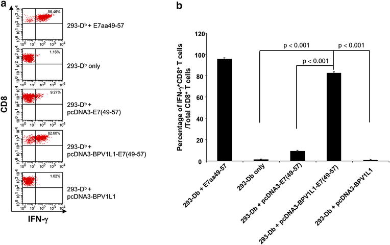
Fig. 2.

Presentation of HPV16 E7 antigen by 293-Db cells transfected with various plasmid constructs. HPV16-E7aa49-57 peptide loaded or pcDNA3-E7(49-57), pcDNA3-BPVL1-E7(49-57) or pcDNA3-BPVL1 transfected 293-Db cells were co-incubated with HPV16 E7aa49-47 peptide-specific CD8+ T cell line (E:T ratio at 1:1). Untreated 293-Db cells were used as negative control. After co-incubation, the E7-specific CD8+ T cells were stained for surface CD8 and intracellular IFN-γ and acquired with FACSCalibur flow cytometer. The data was analyzed using CellQuest software. a Representative flow cytometry analysis. b Bar graph summary of the flow analysis
