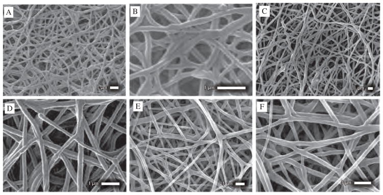
Figure 4.
SEM images of (A), (B) NFs set D, category 3 at different magnification and location (C), (D) NFs set E, category 3 at different magnification and location showing uniform NFs formation without large beads or drug nanocrystals outside of sheath (E), (F) NFs set F, category 3 at different magnification and location showing uniform, thin NFs formation without large beads or drug nanocrystals outside of sheath.

