
Figure 2.
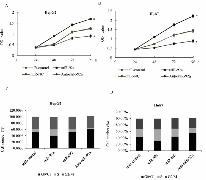
miR-92a promoted cell growth and proliferation of hepatocellular carcinoma (HCC) cells
A) MTT assay was performed in HepG2 cells transfected with miR-92a or control mimics, or transfected with anti-miR-92a or miR-NC. Optical density (OD) was measured 12, 24, 36, 48, 72, and 96 hr after transfection. B) MTT assay was performed in the Huh7 cells. C) Cell cycle distribution was performed using flowcytometry (FCM) in HepG2 cells transfected with anti-miR-92a or miR-NC, or miR-92a or control mimics. The percentage of cells in the G0/G1, S, and G2/M phase was shown, * P<0.05. D) Cell cycle distribution was performed in Huh7 cells