

Abstract
Objective(s):
Cucurbita moschata Duchesne (pumpkin) is a well-known plant with several pharmacological effects. The aim of the present study was to assess burn wound healing activity of C. moschata peel extract (CE). Also, standardized CE was assessed for antioxidant activity and antibacterial effects against major pathogens of burns.
Materials and Methods:
Healing properties of topical preparation of 10% and 20% concentrations of CE were assessed on second degree burn in rats during a 14-day period as well as histological studies, total antioxidant power, lipid peroxidation and total thiol content of skin tissue samples.
Results:
Radical scavenging IC50 and ferric-reducing antioxidant power value were 4.015±0.20 mg/ml and 142.63±2.65 mmol Fe2+/g, respectively. Total mucilage content was 13.8%. The optimal results were obtained by 20% CE that showed 90.80±5.86 % wound closure and tissue repair as well as significant reduction of tissue oxidative stress biomarkers. Histological analyses confirmed wound healing activity of pumpkin peel extract.
Conclusion:
Considering the high mucilage content of the plant, providing a moist environment for wound, C. moschata peel extract could be a natural remedy for treatment of burns. Further clinical studies are suggested to confirm C. moschata peel extract as a wound healing agent.
Keywords: Cucurbita moschata, Histology, Traditional persian medicine, Pumpkin, Wound healing
Introduction
Burn injury is defined as skin damage caused by extreme heat, radiation, electricity or corrosive chemicals. Based on the depth, burn wounds are classified into superficial (first degree), partial thickness (second degree) and full thickness (third degree). The process of burn wound healing consists of five main phases: 1) Inflammation, in which the chemoattractant mediators attract immune cells to the wound area, 2) re-epithelialization which starts with the proliferation and migration of epithelial cells at the wound edge, probably due to lack of neighbor cells, 3) granulation with the production of extracellular matrix by fibroblasts, 4) neovasculariza-tion which is mostly induced by pro-angiogenic factors (mainly produced by macrophages),-5) and ultimately the last phase which is wound contraction (1, 2).
Epidermal growth factor, fibroblast growth factor family, transforming growth factor β1 and β2, platelet-derived growth factor, vascular endothelial growth factor, tumor necrosis factor α, interleukin-1, insulin-like growth factor-1 and colony-stimulating factor-1 are the cytokines which are involved in the wound healing process (3, 4).
During a traumatic event like burn, free radicals overwhelm natural radical scavengers in the body and reactive oxygen species (ROS) are increased in the damaged tissue. On the other hand, neutrophil activation ramps up the amount of free radicals. Moreover, burn injury partially suppresses the immune system. Hence, the use of antioxidants may have a positive effect on the restoration of skin tissue damage and other burn wound complications (5).
Since skin contains the nonspecific front line immune system components, inappropriate treat-ment of extensive burn injuries can lead to life-threatening infections. There are several conventional treatments for burn wounds such as silver sulfadiazine (as a topical antibacterial agent) or different types of wound dressings; however, there are numerous number of plants which are potentially able to improve the healing process of burn wounds (1, 6).
Medicinal plants used in traditional medicines of different countries are considered as valuable sources for the discovery of new drugs (7, 8). A vast variety of topical formulations including ointments and poultices are mentioned to be useful in burn wound management in the text books of Traditional Persian Medicine (TPM). One of the entries in the texts with burn healing effect is the fruit peel of Cucurbita moschata.
C. moschata Duchesne (synonym Pepo moschata), commonly known as “pumpkin” and belonging to the family Cucurbitaceae, is a widely distributed plant mainly used for its fruit and seeds (9).
Pumpkin fruit is nutritionally rich due to the carotenoid and γ-tocopherol and is demonstrated to have anti-fatigue activity in mice (10). Noor Aziah et al. suggested that peeled and unpeeled pumpkin flour is a good enrichment material for wheat flour due to the rich content of protein, carbohydrate, essential minerals and trace elements (11). Different amino acids including alanine, arginine, aspartic acid, glutamic acid, histidine, leucine, isoleucine, glycine, lysine, methionine, phenylalanine, serine, threonine, valine and tyrosine have been detected in pumpkin peel.
Pumpkin seed oil contains mono and polyunsaturated fatty acids as well as saturated ones like palmitic acid, stearic acid, oleic acid and linoleic acid (12).
Several pharmacological activities have been proposed for different parts of this plant. Common and sugar-removed powder of the pumpkin fruit reduced blood glucose and increased plasma insulin (13, 14). Crude extract of pumpkin fruit showed antidiabetic activity in alloxan-induced diabetic mice (15). Pumpkin powder also reduced serum total cholesterol and triglyceride in diabetic animals (16).
In TPM, pumpkin peel was used for the treatment of hepatic disorders, peptic ulcer, gastrointestinal bleeding and different types of wounds, including burn wound. According to TPM text books such as “Makhzan –al-Adviah” by M.H. Aghili, pimpkin fruit is considered to have cool and wet nature and can be used in hot and dry diseases including burn wounds, which have a hot nature. Additionally, some preparations of the fruit peel are suggested to be useful for burn wound healing (17).
To the best of our knowledge, there is no study on the burn wound healing activities of C. moschata fruit peel; thus, the aim of the present study is to assess burn wound healing properties of C. moschata peel extract based on TPM knowledge.
Materials and Methods
Chemicals
1,1-diphenyl-2-picrylhidrazyl (DPPH), butylated hydroxy anisole (BHA), 2,4,6-tri(2-pyridyl-s-triazine) or TPTZ, FeCl3.6H2O, Folin-Ciocalteu and pentobarbital from Sigma Chemical Company (USA), gallic acid, sodium bicarbonate, acetic acid, acetic anhydride, hydro- chloric acid (HCl), ethanol, methanol, dimethyl sulfoxide (DMSO), Mueller-Hinton agar, sodium chloride, potassium dihydrogen phosphate, dipotassium hydrogen phosphate, phosphoric acid, 2-thiobarbituric acid (TBA), trichloroacetic acid, H2SO4, n-butanol, and 5’5’dithio-bisnitrobenzoic acid (DTNB) from Merck company (Germany) and silversulfadiazine 1% cream (Behvazan, Iran) were purchased for this study.
Plant material standardization
Pumpkin fruits were collected from Varamin, South west of Tehran, Iran in October 2013. The plant was authenticated by Professor Gh. Amin and the voucher specimen was deposited under voucher number: 6759-TEH in the Herbarium of Faculty of Pharmacy, Tehran University of Medical Sciences, Tehran, Iran.
Pumpkin fruits were peeled and dried at room temperature. 150 g of dried peel was ground into coarse powder and soaked in 70% ethanol in a percolator apparatus. The extraction was performed every 24 hr for three times and the extracts were mixed together. The extract was concentrated with a rotary evaporator at 40° C until a viscous liquid was obtained. Moisture content, total ash, water soluble ash and acid insoluble ash of dried peel were measured according to WHO guidelines (18). All experiments were done in triplicates.
DPPH radical scavenging activity
DPPH radical scavenging activity was carried out by the following method: 2 ml of DPPH methanolic solution (0.04 mg/ml) was mixed with 1 ml of different concentrations of pumpkin extract (1, 2, 4 and 6 mg/ml) or BHA (0.001%) as reference standard. The solutions were kept in dark for 45 min until the absorbance was measured at 517 nm with UV/visible spectrophotometer (Mecasys, Korea). To eliminate the intrinsic absorption of the extract and solvent, ethanol and a solution of 1 ml of extract with 2 ml of methanol were used as blank and negative control, respectively. The radical scavenging activity was calculated using the below formula:
In which sample absorbance is defined as the absorbance of different extract concentrations mixed with DPPH, blank absorbance is the absorbance of DPPH with ethanol without any extract and control absorbance is the absorbance of the mixture of 1 ml of extract and 2 ml of methanol (19).
Total antioxidant capacity
Ferric-reducing antioxidant power (FRAP) was determined by the following method: 1.5 ml of fresh FRAP reagent (25 ml of 0.3 M acetate buffer, 2.5 ml of TPTZ solution, and 2.5 ml of FeCl3 6H2O solution) was mixed with 50 µl of diluted extract and after 10 min the absorbance was recorded at 593 nm (19).
Total phenol assay
To estimate the total phenolic content, 1 ml of a 1 mg/ml of the extract in ethanol was mixed with 1.5 ml of fresh 10% v/v Folin-Ciocalteu reagent and after 5 min, 1.5 ml of 6% w/v sodium bicarbonate was added to the solution. After 90 min, the absorbance was measured at 765 nm. Gallic acid was used as standard and total phenolics content was expressed as gallic acid equivalent (20).
Total mucilage content
Mucilage content was estimated by the modified Kalyansundaram method. 1 g of the plant powder was added to 10 ml of boiling 0.1 N HCl. The mixture was heated for 5 min (the optimum time for mucilage extraction was achieved experimentally), then was filtered through clean muslin cloth before becoming cold. The residual was washed twice with 5 ml of hot water and the final filtrate was mixed with 60 ml of 96% ethanol. The mixture was then allowed to stand at 4 °C refrigerator. After 5 hr, the supernatant fluid was decanted off, the precipitate was dried in the oven over night at 50 °C and was weighted (21, 22).
In vitro antibacterial activity
The antibacterial effect of the pumpkin extract was assessed using the conventional agar dilution method. The test microorganisms were Staphylococcus aureus ATCC 6538 and Pseudomonas aeruginosa ATCC 9027. Two-fold dilution of test materials and ciprofloxacin were prepared in 1 ml of DMSO. Each sample was added to 14 ml of molten Mueller-Hinton agar to reach the final concentrations of 30-0.47 mg/ml for test material and 100-0.0031 µg/ml for ciprofloxacin. The microbial suspensions were prepared by suspending 24 hr cultures from Mueller-Hinton agar media in 0.9% saline and were adjusted photometrically at 600 nm to a cell density equal to about 0.5 McFarland standard (1.5×10⁸ CFU/ml). The bacterial inocula were then diluted in 0.9% saline to achieve 107 CFU/ml. The plates were spot-inoculated with 3 µl of each bacterial suspension (final inoculation: 104 CFU/ml). Minimum inhibitory concentration (MIC) was determined after 24 hr incubation at 37 °C as the lowest concentration of each test material inhibiting the visible growth of microorganisms on the plate (23).
Ointment preparation
Two concentrations (10% and 20%) of Cucurbita extract (CE) was prepared in eucerine base. Silver sulfadiazine cream 1% and eucerine were used as positive and negative controls, respectively.
In vivo assessment of burn wound healing activity
Total of 24 male albino Wistar rats (6 in each group) of weight ranging 200-250 g were used. The rats were housed in standard vivarium condition (temperature of 25±1 °C and relative humidity of 60%, 12 hr light and 12 hr dark cycles) and were fed with standard laboratory food and water ad libitum. The experiment was approved by the animal ethics guidelines of Tehran University of Medical Sciences.
Animals were shaved at the dorsal part under anesthesia with intra-peritoneal injection of pento-barbital (50 mg/kg) and second degree burn wounds were created using an electrical heater with a circular probe (radius of 1 cm and 110 °C heat for 10 sec). Experimental animals were kept individually (one in each cage) and were randomly divided into 4 groups receiving: 10% CE, 20% CE, silver sulfadiazine1% and eucerine starting 24 hr after burn induction. Ointments were applied topically to cover the wound area every 24 hr for 14 days. Wound closure process was monitored by taking photographs using a paper ruler as scale and the closure rate was estimated as the percentage decrease in wound size with Adobe Photoshop CS3. Wound closure percentage was measured using the following formula:
Wound closure (%) = 100× [(first day wound size – specific wound size) / first day wound size]
The rats were sacrificed on the day 14 and the collected skin samples of wound area were cut in halves. One half was kept in -80 °C for measurement of lipid peroxidation (LPO), total antioxidant power (TAP) and total thiol molecules (TTM). For this purpose, defrosted tissues were immersed in phosphate-buffered saline and were homogenized using Heidolph Silent Crusher M (Germany). The samples were centrifuged at 3000 g for 15 min in 4 °C and the supernatant fluid was used for further analyses (24, 25).
Histological analyses
The other part of the wound area tissue samples was preserved in buffered formalin 10% to assess the histological changes. Tissue sections were stained with hematoxylin and eosin and microscopic photographs were captured under × 400 magnifications (24).
Tissue biomarkers of oxidative stress
LPO
A solution of 20% trichloroacetic acid was mixed with tissue extract and the precipitant was reacted with H2SO4 (0.05 M). 2-Thiobarbituric acid (TBA, 0.2% in 2 M sodium sulfate) was then added and after 30 min heating in boiling water bath, the mixture was extracted with n-butanol and absorbance was measured at 532 nm (ELISA reader, Biotek, Germany) (25).
TAP
For determination of total antioxidant power of samples, tissue extracts were mixed with fresh FRAP reagent (25 ml of 0.3 M acetate buffer, 2.5 ml of TPTZ solution, and 2.5 ml of FeCl3 6H2O solution) and absorbance was measured at 593 nm (25, 26).
TTM
To assess total thiol content, tissue extract was mixed with Tris- EDTA buffer (Tris base 0.25 M, EDTA 20 mM, pH: 8.2) and the reaction of DTNB with total sulfhydryl groups was used to reach a chromogen material and the absorbance was measured at 420 nm (25, 27).
Statistical analyses
All data analyses were carried out using the SPSS 15 software (one-way ANOVA and Tukey`s Post hoc test). P value of less than 0.05 was considered as statistical significance (24).
Results
Plant material standardization
The yield of extraction was 52.13%. Moisture content, total ash, water soluble ash and acid insoluble ash are represented in Table 1.
Table 1.
Moisture and ash content of Cucurbita moschata powder
| Moisture content (w/w%) | Total ash (w/w%) | Water soluble ash (w/w%) | Acid insoluble ash (w/w%) |
|---|---|---|---|
| 90.76±0.16 | 7.76±0.24 | 3.22 ± 1.37 | 2.48 ± 0.88 |
DPPH radical scavenging activity
The IC50 value for DPPH radical scavenging activity was found to be 4.015 ± 0.20 mg/ml (Table 2).
Table 2.
Antioxidant properties and quantitative phytochemical analyses of Cucurbita moschata extract
| DPPH IC50 (mg/ml) | FRAP (mmol Fe2+/g) | Total phenol (mg of gallic acid equivalent/g extract) | Total mucilage (%w/w) |
|---|---|---|---|
| 4.015±0.20 | 142.63±2.65 | 22.92±1.06 | 13.8 |
DPPH: 1, 1-diphenyl-2-picrylhidrazyl, IC50: inhibitory concentration 50%, FRAP: Ferric-reducing antioxidant power
Total antioxidant capacity
Ferric reduction capability of the extract was 142.63±2.65 mmol Fe2+/g which is an indicator for single electron transfer reaction (Table 2). The references standard BHA showed an IC50 value of 7.2 μg/ml.
Total phenol assay
The total phenolic content of pumpkin peel extract was determined by Folin–Ciocalteu method which was equal to 22.92±1.06 mg of gallic acid in each g of dried extract (Table 2).
Total mucilage content
Total mucilage content of the CP was calculated by the technique of Kalyansundaram which was equal to 13.8% w/w (Table 2).
In vitro antibacterial activity
All the plates containing pumpkin peel extract showed visible growth of both microorganisms which indicated that none of the concentrations could inhibit the growth of test bacteria at 30 mg/ml. Therefore, the MICs of test materials against S. aureus and P. aeruginosa may be higher than 30 mg/ml.
In vivo assessment of burn wound healing activity
Percentage of wound contraction rate is shown in Table 3. There was no significant difference between treated groups before the 10th day. 20% CE showed a significant reduction of wound area on day 10 (P<0.05). On the last day of the experiment (day 14) all treated groups indicated significant closure of wound in comparison to negative control (P<0.01). In addition, wound contraction rate with 20% CE was significantly higher than positive control group (P<0.05).
Table 3.
Wound contraction rate (%)
| Treatment | 10th day | 14th day |
|---|---|---|
| Negative control | 24.67±5.39 | 57.80±5.71 |
| Positive control | 46.17±5.98 | 78.80±3.96**a |
| 10% CE | 29.60±5.54 | 77.60±5.41**a |
| 20% CE | 61.50±6.28*a | 90.80±5.86**a,b |
Values are expressed as mean ± standard deviation for each group. Significant differences:
: P< 0.05,
: P<0.01.
X: in comparison to negative control,
X: in comparison to positive control.
CE: Cucurbita moschata extract
Tissue biomarkers of oxidative stress
LPO
Treatment with 10% and 20% CE significantly decreased lipid peroxidation in skin tissue samples in comparison to negative control (P<0.001, Figure 1).
Figure 1.
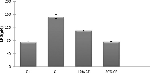
Effect of treatments on lipid peroxidation in skin tissue samples of the day 14
LPO: lipid peroxidation, C-: negative control, C+: positive control, CE: Cucurbita moschata extract. *: P< 0.05, **: P<0.01, ***:P< 0.001
TAP
In TAP test, only 20% CE was able to increase total antioxidant capacity of the damaged tissues (P<0.01, Figure 2).
Figure 2.
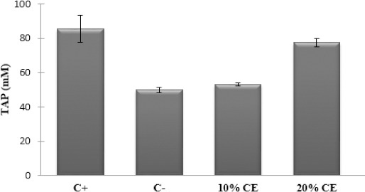
Effect of treatments on total antioxidant power of skin tissue samples of the day 14
TAP: total antioxidant power, C-: negative control, C+: positive control, CE: Cucurbita moschata extract. *: P<0.05, **: P<0.01, ***:P< 0.001
TTM
Significant change in total thiol content of the skin samples was observed with 20% concentrations of CE (P<0.01), but not with 10% ointment (Figure 3).
Figure 3.
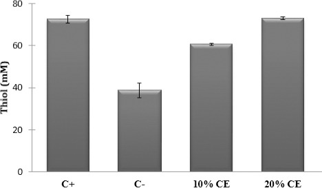
Effect of treatments on total thiol content of skin tissue samples of the day 14
TTM: total thiol molecules. C-: negative control, C+: positive control, CE: Cucurbita moschata extract. *: P< 0.05, **: P<0.01, ***:P< 0.001
Histological analyses
Last day tissue samples of wound area were compared under ×400 magnification and the images are shown in Figure 4.
Figure 4.
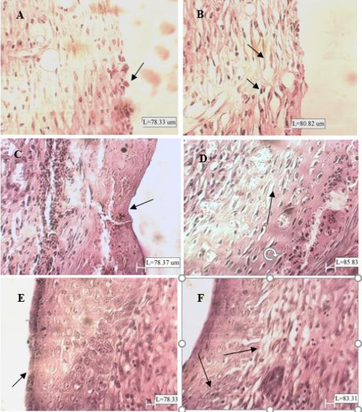
Microscopic images of skin tissue samples of the day 14 with magnification of ×400
A: Re-epithelialization was not observed in the wound center and PMNs are visible in burn wound area. B: Basal layers of epithelium are formed in wound margins, fibroblasts and irregular deposition of collagen bundles are visible. C, D: New epithelium is not formed in the wound center and the there is an irregular arrangement of collagen bundles. E, F: absence of PMN is visible and there is well- organized arrangement of collagen fibers. Formation of epidermis and dermis almost reached the normal level but stratum keratinosum is not regenerated. Cell proliferation in the basal layer of epithelium could be seen. A: negative control, B: positive control, C and D: CE10%, E and F: CE20%. CE: Cucurbita moschata extract, PMN: polymorphonuclear
In control group, there was no epidermal regeneration in the wound center and polymorphonuclears (PMNs) were visible in wound area. Silver sulfadia-zine treated group showed formation of epithelium in wound margins and irregular deposition of collagen bundles. In 10% CE, new epithelium was not formed in the wound center and there was an irregular deposition of collagen bundles. 20% CE treated animals showed absence of PMNs and well-organized arrangement of collagen fibers. Although the stratum keratinosum was not regenerated, formation of different layer of skin tissue and cell proliferation in the basal layer of epithelium almost reached the normal level. Newly formed epidermis and dermis indicated better wound healing activity of 20% CE in comparison to negative and positive control groups.
Discussion
Burn wound healing is an orchestrated cascade of damaged skin cells replacement which results in tissue repair. Several chemokines and cytokines are involved in this process as well as immune cells including neutrophils, monocytes and macrophages (3, 4). In the present study, for the first time, an evaluation of burn wound healing properties of pumpkin peel and its antibacterial effect on predominant pathogens of burn wounds was performed. Raw material was evaluated by measuring the moisture of the fresh peel, total ash, water soluble ash and acid insoluble ash content of the dried peel; however, it should be mentioned that as pumpkin fruit peel has not been previously considered as a useful plant material for medicinal applications, no specific standard for the moisture and ash content is currently available. Thus, our measurement, as the first reported data on pumpkin fruit peel, can be used in future studies as parameters for primary quality control of the plant material. The fruit peel extract was also standardized based on the total phenolic compounds since this category of phyto-chemicals was supposed to play an important role in wound healing activity of the extract. Total mucilage content was also evaluated since numerous number of plants with high mucilage content like quince seed and Malva spp. showed anti-inflammatory and wound healing effects (28, 29).
Plants and their large number of phytochemical constituents like phenolic compounds, flavonoids, anthocyanins and tannins with their astringent, anti-inflammatory and antioxidant effects are demons-trated to be effective in different types of pathologic conditions like wounds (30-32). Previous phytochemical screening tests demonstrated the presence of flavonoids and phenolic compounds in both methanolic and ethanolic extract of C. moschata peel (33).
According to previous publications, Pumpkin extract enhanced antioxidant enzymes e.g. super oxide dismutase (SOD) and glutathione peroxidase (GSH-Px) activities and suppressed lipid peroxidation in mice compared with placebo treated group (34). It also improved the proliferation of splenic lymphocytes and natural killer cells which shows an immunomodulatory activity for the plant (35). Pumpkin oil demonstrated antibacterial activity against a wide range of bacteria. There are also some reports on anticancer, antimutagenic and anthelmintic properties of pumpkin (9, 36). DPPH radical scavenging activity and total antioxidant power of the extract also suggest an antioxidant mechanism of burn wound healing which was confirmed by tissue evaluations of oxidative damage. A previous study also demonstrated the antioxidant properties of pumpkin fruit extract and seed oil (37). When administered systemically, pumpkin extract decreased malonaldehyde (MDA) and elevated liver SOD and GSH-px (35). It is the first time that total antioxidant power and radical scavenging activity of pumpkin fruit peel was assessed.
Pumpkin peel could not show significant anti-bacterial activity on wound pathogens including S. aureus and P. aeruginosa. A study on antibacterial effects of C. pepo, another species of the pumpkin genus, reported inhibition zone of 6-10 mm with 0.5-2 mg/disc of ethanolic and methanolic peel extract against S. aureus. However, differences in the species of the plant and antibacterial evaluation method along with not reporting the ATCC number of the microorganism as well as lack of a gold standard antibacterial agent as positive control make our investigation not be comparable to this study (33).
Topical 20% CE showed the best in vivo result amongst all test materials. In addition to the better result compared with control, significant increase in wound closure percentage was observed in 20% CE treated group compared to that of positive control group. Wound areas showed fully developed epithelialization representing cell proliferation and migration, especially in 20% CE treated group.
Regarding the oxidative stress biomarkers, 20% CE and 10% CE treated groups showed a lower degree of lipid peroxidation as well as higher level of total antioxidant capacity. Total thiol molecules, one of the direct targets of oxidant signals, were also significantly elevated, especially in 20% CE treated group, compared with control. Despite the relatively high IC50 and FRAP value of the extract, results of the tissue biomarkers of oxidative stress-TAP, LPO and TTM which provides more valuable data in comparison to in vitro antioxidant assessments- demonstrates that antioxidant phytochemicals of the extract could adequately penetrate into the damaged tissue and represent in vivo antioxidant activity.
Total mucilage content of pumpkin peel was measured as 13.8% w/w which is relatively high in comparison to well-known mucilaginous herbs like species from Malvaceae or Plantaginaceae families (38, 39). There is a growing body of evidence which demonstrates a better healing of wounds in a moist environment. An occlusive dressing results in a faster re-epithelialization, autolythic debridement, angiogenesis, better migration of keratinocytes as well as induction of hypoxia inducible factor-1 which causes an increase in the production of endogenous wound healing stimulants (40). High mucilage content of pumpkin peel can provide a suitable moist environment for damaged skin cells which finally results in a better burn wound healing.
Conclusion
According to the aforementioned results, it is suggested that the pumpkin peel extract could act as a burn healing agent mainly via its moisturizing effect as well as elevation of tissue antioxidant power. Based on the previous studies, other mechanisms are also proposed for wound healing effect of the plant which can be assessed in future studies. Investigations have proven the immunomodulatory effect of pumpkin by promotion of lymphocytes proliferation and natural killer cells activity as well as enhancement of CD4+ and CD8+ cells which can be suitable targets for future studies on the mechanism of wound healing activity of the plant (9).
Conclusion
Results obtained from current study showed beneficial effect of C. moschata peel extract, especially with 20% concentration, in animal model of burn wound through providing a moist environment due to its high content of mucilage, reduction of tissue oxidative stress biomarkers and histopathological condition of damaged tissue. Further investigations are needed for more definite confirmation of wound healing activity of pumpkin peel.
Acknowledgment
This study is a part of a Pharm D thesis and a project which has been funded and supported by Tehran University of Medical Sciences (TUMS).
References
- 1.Bahramsoltani R, Farzaei MH, Rahimi R. Medicinal plants and their natural components as future drugs for the treatment of burn wounds: an integrative review. Arch Dermatol Res. 2014;306:601–617. doi: 10.1007/s00403-014-1474-6. [DOI] [PubMed] [Google Scholar]
- 2.Wu F, Bian D, Xia Y, Gong Z, Tan Q, Chen J, et al. Identification of major active ingredients responsible for burn wound healing of centella asiatica Herbs. Evid Based Complement Alternat Med. 2012;2012:848093. doi: 10.1155/2012/848093. [DOI] [PMC free article] [PubMed] [Google Scholar]
- 3.Singer AJ, Clark RA. Cutaneous wound healing. N Engl J Med. 1999;341:738–746. doi: 10.1056/NEJM199909023411006. [DOI] [PubMed] [Google Scholar]
- 4.Ching YH, Sutton TL, Pierpont YN, Robson MC, Payne WG. The use of growth factors and other humoral agents to accelerate and enhance burn wound healing. Eplasty. 2011;11e41:429–449. [PMC free article] [PubMed] [Google Scholar]
- 5.Sahib AS, Al-Jawad FH, Alkaisy AA. Effect of antioxidants on the incidence of wound infection in burn patients. Ann Burns Fire Disasters. 2010;23:199–205. [PMC free article] [PubMed] [Google Scholar]
- 6.Kumar B, Vijayakumar M, Govindarajan R, Pushpangadan P. Ethnopharmacological approaches to wound healing-exploring medicinal plants of India. J Ethnopharmacol. 2007;114:103–113. doi: 10.1016/j.jep.2007.08.010. [DOI] [PubMed] [Google Scholar]
- 7.Rahimi R, Baghaei A, Baeeri M, Amin G, Shams-Ardekani MR, Khanavi M, et al. Promising effect of Magliasa, a traditional Iranian formula, on experimental colitis on the basis of biochemical and cellular findings. World J Gastroenterol. 2013;19:1901–1911. doi: 10.3748/wjg.v19.i12.1901. [DOI] [PMC free article] [PubMed] [Google Scholar]
- 8.Farzaei MH, Ghasemi-Niri SF, Abdolghafari AH, Baeeri M, Khanavi M, Navaei-Nigjeh M, et al. Biochemical and histopathological evidence on the beneficial effects of Tragopogon graminifolius in TNBS-induced colitis. Pharm Biol. 2015;53:429–436. doi: 10.3109/13880209.2014.923004. [DOI] [PubMed] [Google Scholar]
- 9.Caili F, Huan S, Quanhon L. A review on pharmacological activities and utilization technologies of pumpkin. Plant Foods Hum Nutr. 2006;61:73–80. doi: 10.1007/s11130-006-0016-6. [DOI] [PubMed] [Google Scholar]
- 10.Wang SY, Huang WC, Liu CC, Wang MF, Ho CS, Huang WP, et al. Pumpkin (Cucurbita moschata) fruit extract improves physical fatigue and exercise performance in mice. Molecules. 2012;17:11864–11876. doi: 10.3390/molecules171011864. [DOI] [PMC free article] [PubMed] [Google Scholar]
- 11.Noor Aziah AA, Komathi CA. Physicochemical and functional properties of peeled and unpeeled pumpkin flour. J Food Sci. 2009;74:S328–333. doi: 10.1111/j.1750-3841.2009.01298.x. [DOI] [PubMed] [Google Scholar]
- 12.Kim MY, Kim EJ, Kim YN, Choi C, Lee BH. Comparison of the chemical compositions and nutritive values of various pumpkin (Cucurbitacea) species and parts. Nutr Res Pract. 2012;6:21–27. doi: 10.4162/nrp.2012.6.1.21. [DOI] [PMC free article] [PubMed] [Google Scholar]
- 13.Ju LY, Chang D. Hypoglycemic effect of pumpkin powder. J Harbin Med. 2001;21:5–6. [Google Scholar]
- 14.Chen JG. Effects of sugar-removed pumpkin zymptic powders in preventing and treating the increase of blood glucose in alloxan-induced diabetic mice. Chin J Clin Rehabil. 2005;9:94–95. [Google Scholar]
- 15.Jin H, Zhang YJ, Xiang JX, Zhu LY, Chen P, Li J, Yao HY. Studies on the extraction of pumpkin components and their biological effects on blood glucose of diabetic mice. J Food Drug Anal. 2013;21:184–189. [Google Scholar]
- 16.Zhang ZJ. Effects of superfine pumpkin powder on alloxan induced Diabetes Mellitus rabbits. J Chin Cereals Oils Assoc. 1998;13:52–56. [Google Scholar]
- 17.Aghili M. In: Makhzan-al-Advia 1771. Rahimi R, Shams Ardekani MR, Farjadmand F, editors. Tehran: University of Medical Sciences Tehran; 2009. [Google Scholar]
- 18.World Health Organization. Quality control methods for herbal materials. 2011 [Google Scholar]
- 19.Farzaei MH, Rahimi R, Attar F, Siavoshi F, Saniee P, Hadjimahmoodi M, et al. Chemical composition, antioxidant and antimicrobial activity of essential oil and extracts of Tragopogon graminifolius, a medicinal herb from Iran. Nat Prod Commun. 2014;9:121–124. [PubMed] [Google Scholar]
- 20.Farzaei MH, Khanavi M, Moghaddam G, Dolatshahi F, Rahimi R, Shams-Ardekani MR, et al. Standardization of Tragopogon graminifolius DC. extracts based on phenolic compounds and antioxidant activity. J Chem. 2014 doi:10.1155/2014/425965. [Google Scholar]
- 21.Mirmasumi M, Ebrahimzadeh H, Tabatabaei MF. Mucilage production in tissue culture of plantago lanceolata. J Agric Sci Technol. 2001;3:155–160. [Google Scholar]
- 22.Kalyansundaram NK, Amin DR, Dalal KC. In: “Biannual Report (from Oct. 1978 to Nov. 1980) of All India Coordinated Project on Medicinal and Aromatic Plants. Gujarat Agric. Unive., Anand; 1980. Quality Evaluation of Isabgol Seeds; pp. 125–127. [Google Scholar]
- 23.Seghatoleslami S, Samadi N, Salehnia A, Azimi S. Antibacterial activity of endemic Satureja Khuzistanica Jamzad essential oil against oral pathogens. Iran Endod J. 2009;4:5–9. [PMC free article] [PubMed] [Google Scholar]
- 24.Kahkeshani N, Farahanikia B, Mahdaviani P, Abdolghaffari A, Hassanzadeh G, Abdollahi M, et al. Antioxidant and burn healing potential of Galium odoratum extracts. Res Pharm Sci. 2013;8:197–203. [PMC free article] [PubMed] [Google Scholar]
- 25.Navaei-Nigjeh M, Rahimifard M, Pourkhalili N, Nili-Ahmadabadi A, Pakzad M, Baeeri M, Abdollahi M. Multi-organ protective effects of cerium oxide nanoparticle/selenium in diabetic rats: evidence for more efficiency of nanocerium in comparison to metal form of cerium. Asian J Anim Vet Adv. 2012;7:605–612. [Google Scholar]
- 26.Abdolghaffari AH, Baghaei A, Moayer F, Esmaily H, Baeeri M, Monsef-Esfahani HR, et al. On the benefit of Teucrium in murine colitis through improvement of toxic inflammatory mediators. Hum Exp Toxicol. 2010;29:287–295. doi: 10.1177/0960327110361754. [DOI] [PubMed] [Google Scholar]
- 27.Sethi J, Sood S, Seth S, Talwar A. Evaluation of hypoglycemic and antioxidant effect of Ocimum sanctum. Indian J Clin Biochem. 2004;19:152–155. doi: 10.1007/BF02894276. [DOI] [PMC free article] [PubMed] [Google Scholar]
- 28.Kovalik AC, Bisetto P, Pochapski MT, Campagnoli EB, Pilatti GL, Santos FA. Effects of an orabase formulation with ethanolic extract of Malva sylvestris L. in oral wound healing in rats. J Med Food. 2014;17:618–624. doi: 10.1089/jmf.2013.0001. [DOI] [PubMed] [Google Scholar]
- 29.Tamri P, Hemmati A, Boroujerdnia MG. Wound healing properties of quince seed mucilage in vivo evaluation in rabbit full-thickness wound model. Int J Surg. 2014;12:843–847. doi: 10.1016/j.ijsu.2014.06.016. [DOI] [PubMed] [Google Scholar]
- 30.Farzaei MH, Abbasabadi Z, Shams-Ardekani MR, Abdollahi M, Rahimi R. A comprehensive review of plants and their active constituents with wound healing activity in traditional Iranian medicine. Wounds. 2014;26:197–206. [PubMed] [Google Scholar]
- 31.Farzaei MH, Rahimi R, Abdollahi M, Abbasabadi Z. An evidence-based review on medicinal plants used for the treatment of peptic ulcer in traditional Iranian medicine. Int J Pharmacol. 2013;9:108–124. [Google Scholar]
- 32.Rahimi R, Mozaffari S, Abdollahi M. On the use of herbal medicines in management of inflammatory bowel diseases: a systematic review of animal and human studies. Dig Dis Sci. 2009;54:471–480. doi: 10.1007/s10620-008-0368-x. [DOI] [PubMed] [Google Scholar]
- 33.Chonoko U, Rufai A. Phytochemical screening and antibacterial activity of cucurbita pepo (Pumpkin) against Staphylococcus aureus and Salmonella typhi. Bayero J Pure App Sci. 2011;4:145–147. [Google Scholar]
- 34.Dang C. Effect of pumpkin distillable subject on lipid peroxidation and the activity of antioxidative enzyme induced by Plumbum in mouse. J Clin Rehabil. 2004;8:4378–4379. [Google Scholar]
- 35.Xia HC, Li F, Li Z, Zhang ZC. Purification and characterization of Moschatin, a novel type I ribosome-inactivating protein from the mature seeds of pumpkin (Cucurbita moschata), and preparation of its immunotoxin against human melanoma cells. Cell Res. 2003;13:369–374. doi: 10.1038/sj.cr.7290182. [DOI] [PubMed] [Google Scholar]
- 36.Yadav M, Jain S, Tomar R, Prasad GB, Yadav H. Medicinal and biological potential of pumpkin: an updated review. Nutr Res Rev. 2010;23:184–190. doi: 10.1017/S0954422410000107. [DOI] [PubMed] [Google Scholar]
- 37.Azizah AH, Wee KC, Azizah O, Azizah M. Effect of boiling and stir frying on total phenolics, carotenoids and radical scavenging activity of pumpkin (Cucurbita moschato) Int Food Res J. 2009;16:45–51. [Google Scholar]
- 38.Pakrokh Ghavi P. The extraction process optimization of antioxidant polysaccharides from Marshmallow (Althaea officinalis L.) roots. Int J Biol Macromol 2015. 75:51–57. doi: 10.1016/j.ijbiomac.2014.11.047. [DOI] [PubMed] [Google Scholar]
- 39.Prosky L, Asp NG, Furda I, DeVries JW, Schweizer TF, Harland BF. Determination of total dietary fiber in food and food products: collaborative study. J Assoc Off Anal Chem. 1985;68:677–679. [PubMed] [Google Scholar]
- 40.Broussard KC, Powers JG. Wound dressings: selecting the most appropriate type. Am J Clin Dermatol. 2013;14:449–459. doi: 10.1007/s40257-013-0046-4. [DOI] [PubMed] [Google Scholar]