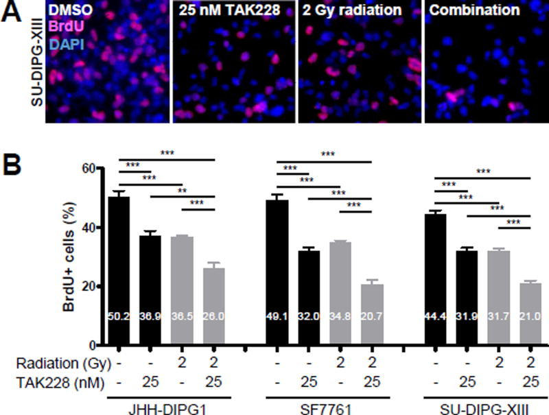
Figure 2.
TAK228 suppressed DIPG proliferation and enhanced radiosensitization
A) Representative 400× photomicrographs showing decreased proliferation as measured by BrdU immunofluorescence (red) after treatment with 25 nM TAK228 for 3 days followed by 2 Gy radiation. DAPI counterstains nuclei (blue). B) Quantification of DIPG proliferation after TAK228 and radiation treatment ***: p <0.0001, **: p <0.01, *: p <0.05 vs. DMSO by t-test.

