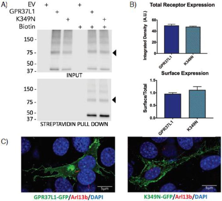
Figure 2. Expression and localization of GPR37L1 wild-type and K349N mutant are equivalent.

(A & B) Transient transfection of Flag-tagged versions of GPR37L1 wild-type or K349N mutant in HEK293T cells yielded comparable levels of receptor expression (upper panels) and trafficking to the cell surface (lower panels). Full-length GPR37L1 runs on SDS-PAGE gels at ~70–75 kDa (arrow head) with higher order bands representing oligomeric forms of the receptor and the ~37 kDa band representing a cleaved form of the receptor. Results are from three independent experiments (± SEM shown). (C) Confocal microscopy analysis of GFP-tagged versions of GPR37L1 wild-type (left panel) and K349N mutant (right panel) revealed a similar pattern of subcellular localization in NIH-3T3 cells, with both receptor variants being predominantly localized to the plasma membrane and neither version of the receptor exhibiting significant ciliary localization. Cilia were labeled using an anti-Arl13b antibody, indicated in red.
