
Fig. 3.
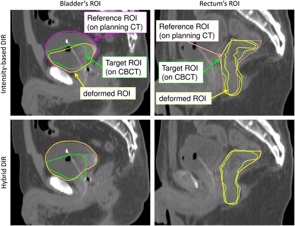
Typical example of rectum and bladder structures based on each DIR (Patient 2). Purple lines: the manual bladder structure on planning CT; green lines: the manual structure on CBCT; yellow lines: the structure deformed by DIR; orange lines: the manual rectum structure on planning CT.