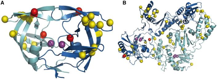
Figure 6.
Structural context of the drug resistance mutations (amino acid replacements) occurring along an example trajectory in HIV-1’s protease protein, PDB code 1KZK (A) and reverse transcriptase protein, PDB code 1RTJ (B), see Fig. 5 for trajectories. The resistance mutations for the protease (M46I and I84V) and reverse transcriptase are coloured red. The catalytic aspartates are coloured purple. The various amino acid replacements occurring prior to the resistance mutation are shown in yellow.

