
Fig. 2.
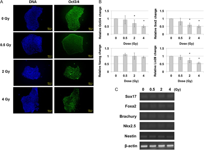
Pluripotency and differentiation marker expressions of hiPS cells 24 h after irradiation. (A) Immunocytochemical analysis of pluripotency marker Oct3/4 in hiPS cells 24 h after irradiation. Green: Oct3/4, Blue: DNA. Scale bar = 100 μm. (B) Real-time-PCR for expression pluripotency markers (Oct3/4, Sox2, Nanog and Lin28) 24 h after irradiation. Each bar represents mean ± s.d. *P < 0.05 (Student's t-test for comparison with non-irradiated cells). (C) Reverse transcription PCR of various differentiation markers of three germ layers. The gene expressions of Sox17, Foxa2 (endoderm marker), Brachury, Nkx2.5 (mesoderm marker) and Nestin (ectoderm marker) were obtained 24 h after irradiation. β-actin served as the loading control.