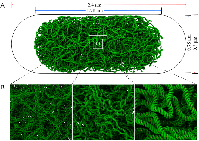
Figure 1.
Structural model of the Escherichia coli chromosome. (A) A model of the 4.6 Mb E. coli chromosome at 1-nt resolution (green) pictured within a volume corresponding to the cell (black outline). Dimensions appearing above and at right are those of the nucleoid (blue) and the cell containing it (red). (B) Images of the chromosome at increasing magnification. In the left panel, individual plectonemes appear; in the right panel, distinct major and minor grooves are visible.

