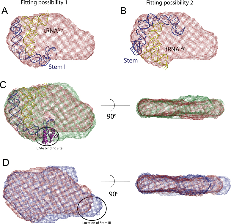
Figure 3.
Location of Stems I and III in the molecular envelope of the Bacillus subtilis glyQS T-box:tRNAGly complex. Panels (A) and (B) show two alternate fittings of the crystal structure of Stem I:tRNAGly complex (12) (PDB ID: 4LCK) in the molecular envelope of the wild-type complex (red). (C) The orientation of Stem I was selected by aligning and comparing the molecular envelopes of the T-box:tRNAGly:L7Ae ternary (green) and wild-type (red) complexes. The 14 kDa L7Ae protein binds to the proximal segment of Stem I and represents a small protrusion in the envelope. A model of the Stem I:tRNAGly crystal structure (12), including the L7Ae structure was placed in the molecular envelope of the T-box:tRNAGly:L7Ae ternary (green), corresponding to the orientation in panel (A). (D) A similar comparison between the envelopes of the wild-type and the T-box (S3.10):tRNAGly complexes helped locate Stem III in the envelope and the model. The diagrams in (C) and (D) show two orthogonal views of the aligned molecular envelopes.

