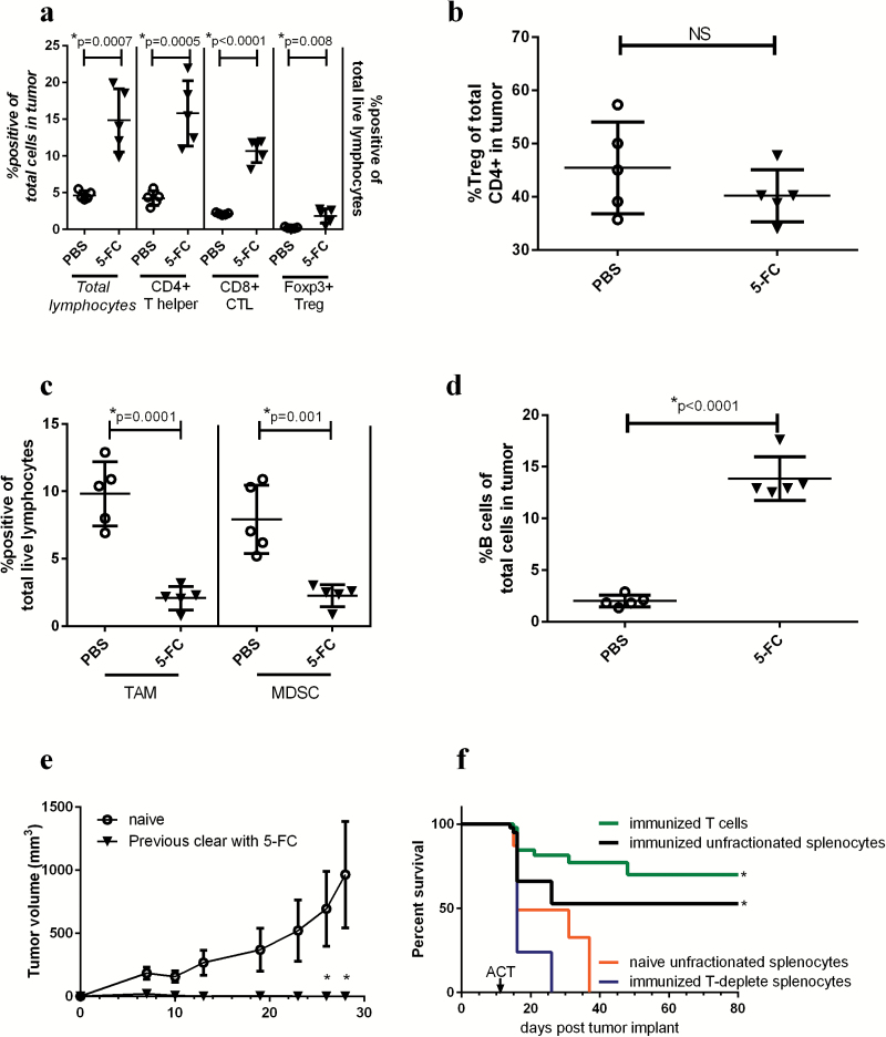
Fig. 4.
Toca 511 and 5-FC treatment continues to reduce immunosuppressive myeloid cells while increasing T cells in the tumor 14 days after treatment initiation. (A) Total lymphocytes were analyzed as a percentage of total cells in the tumor (data correspond to italicized axis on the left), CD4+, CD8+, or Tregs were analyzed as a percentage of total live lymphocytes in the tumor (data correspond to non-italicized axis on the treatment initiation). (B) Tregs were analyzed as a percentage of total CD4+ T cells in the tumor (right) 14 days post 5-FC treatment initiation. (C) TAMs and MDSCs were analyzed in the tumor 14 days after the initiation of 5-FC and expressed as a percentage of total live lymphocytes in the tumor. (D) Fourteen days after 5-FC treatment initiation, B cells were analyzed as a percentage of total live lymphocytes in the tumor. (E) Mice which had cleared tumors through treatment with Toca 511 and 5-FC and were tumor free for 9 weeks were rechallenged with Tu-2449 subcutaneous tumors and tumor burden was assessed over time (n = 5). Age-matched naïve controls were also challenged (n = 10). (F) Animals which had previously cleared Tu-2449 intracranial tumors through treatment with Toca 511 and 5-FC (termed “immunized”) were used for ACT into recipient mice bearing intracranial Tu-2449 tumors. Before transfer, immunized splenocytes were fractionated. Animals received either unfractionated splenocytes from immunized (black) or naïve mice (orange) or purified T cells from immunized spleens (green) or immunized splenocytes depleted of T cells (blue). These data were repeated with similar results in at least one additional experiment. *Statistical significance was defined as P < .05.

