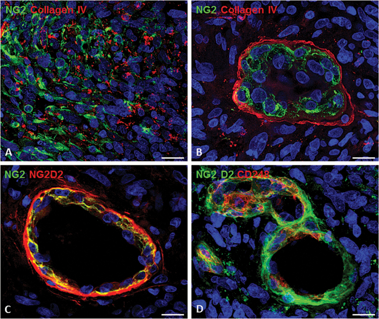
Fig. 2.
The pericyte component of the GBM vessels as shown by confocal microscopy and specific tumoral pericyte markers. (A) NG2-reactive tumoral cells in GBM tissue (collagen type IV in red). (B) A tuft-like vessel (garland vessel; collagen type IV in red), whose multilayered pericyte wall is revealed by a pericyte-specific NG2. Double antibodies against different NG2 isoforms (C) as well as NG2 in double labeling with CD248 (endosialin); (D) pericyte subsets. Bars: (A, C), 25 µm; (B, D), 15 µm. Image from Girolamo F et al. PloS ONE. 2013;8(12):e84883; used with permission.

