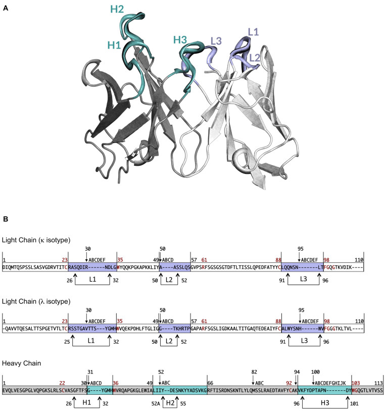
Figure 1.
(A) Variable region of an antibody molecule. Heavy and light chain framework regions are coloured in grey and white, respectively. Loops composing the antigen-binding site are coloured in pale cyan for the heavy chain and light violet for the light chain. (B) Chothia numbering scheme for VH, VK and VL. The numbers above the sequences represent the numbering of specific residues. The remaining residues are numbered consecutively. Letters correspond to insertions. Framework regions are depicted in grey for VH and in white for VK and VL. Complementarity determining regions are coloured in pale cyan for VH and in light violet for VK and VL. Arrows indicate Chothia and Lesk definition of hypervariable loops. Conserved residues are reported in dark red.

