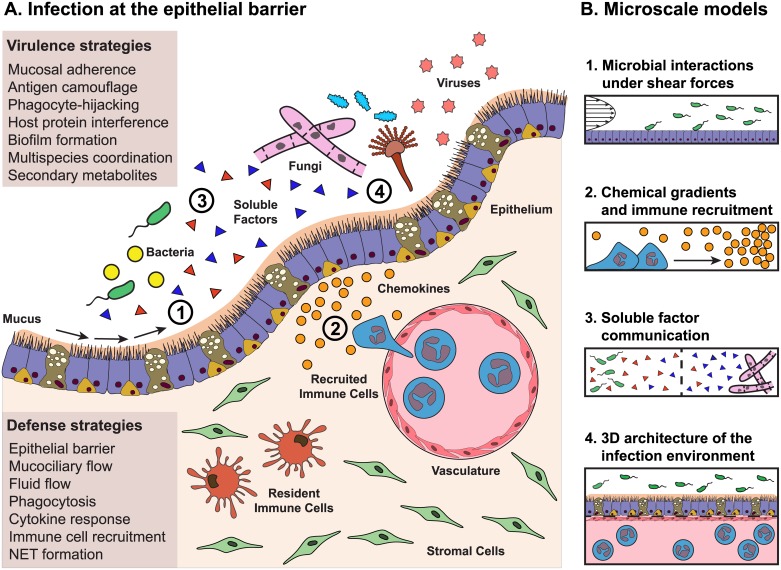
Fig 1. How microscale approaches can be applied to model the complex host pathogen microenvironment.
(A) Virulence strategies of pathogens meet defense strategies of the host at the epithelial barrier. The infectious milieu can be incredibly complex, with organisms from different kingdoms interacting both with each other and with the host. Ideally, an accurate representation of the host, including the epithelium, vascular compartment, interstitial compartment, stromal cells, resident immune cells, and cytokines, would be exposed to an accurate representation of the invading pathogens, including a mixture of bacteria, fungi, viruses, and all sorts of soluble factors. In vitro methods now focus on making this complex environment experimentally tractable, modeling 1 or 2 components of the milieu. (B) Four aspects of the infection microenvironment that can be readily modeled using microfluidic approaches. 1. Microscale techniques simplify the process of modeling the fluid flows and shear forces that occur along the apical surface of the epithelium. 2. Gradients are easy to generate precisely and reproducibly in microscale and are ideal for performing chemotaxis assays. 3. Device geometry is customizable at microscale and offers control over coculture environments, allowing for soluble factor communication between 2 or more populations, as well as coculture between microbes from different kingdoms. 4. Microscale approaches can be used to create organotypic models that recapitulate aspects of tissue structure and function, which may better represent the host in host–pathogen interaction studies. Future microscale approaches may target entirely different aspects of the complex infection microenvironment depicted in (A).

