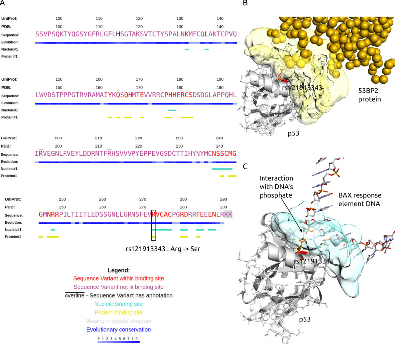
Figure 3.
Summary of GenProBiS results for p53 tumor suppressor protein (gene symbol: TP53; PDB and Chain ID: 1gzhC). (A) Sequence view of p53 with mapped nsSNPs, somatic mis-sense mutations and binding sites. Binding site mis-sense mutation rs121913343 Arg273Ser (red) is located in nucleic and protein–protein binding sites. (B–C) Structural view of p53's (gray cartoon) rs121913343 (red ball-and-sticks) interaction with (B) tumor suppressor p53-binding protein 2 (53BP2) ligand (yellow spheres), where each sphere represents one protein residue and protein binding site on p53 is yellow surface; (C) promoter of proapoptotic gene (Bax) ligand (CPK colored sticks), where the nucleic acid binding site on p53 is a blue surface.

