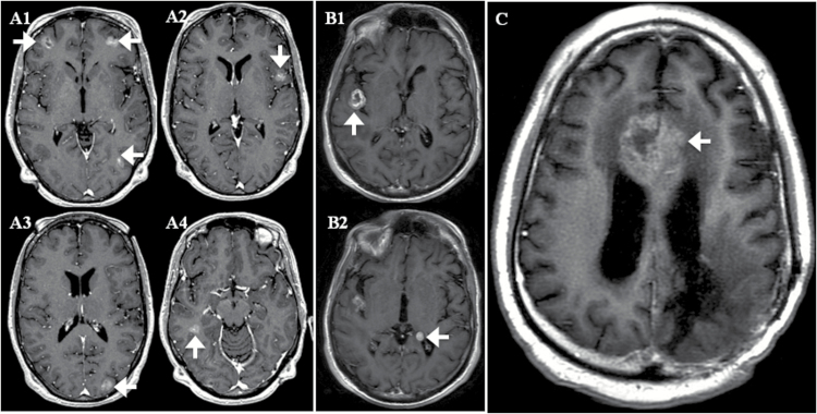
Fig. 3.
MRIs of 3 patients with ≥1 CSF-CTC/mL but negative for LM by study criteria. Axial contrast T1-weighted images of patients (A1–4) and (B1–2) and (C) demonstrating brain parenchymal metastases in close proximity to CSF spaces at the sulci (white arrows) or ventricles (white arrowheads). The 3 patients with breast, bladder, and lung carcinomas had 1, 10, and 64 CSF-CTC/mL, respectively.

