Fig. 1.
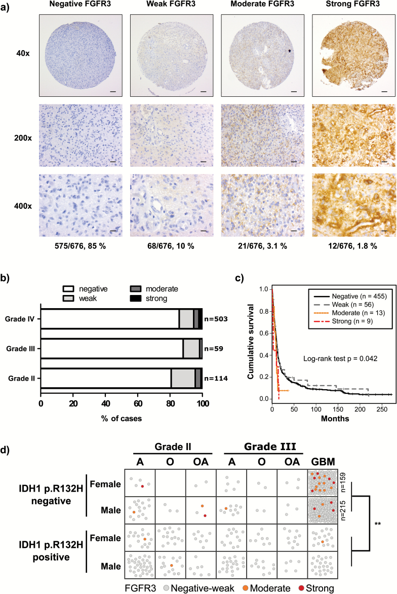
Moderate or strong FGFR3 staining was associated with worse survival and lack of IDH1 p.R132H mutation. (A) Representative staining images. Proportion of samples for each staining category in grades II–IV astrocytomas is indicated below the representative images. Scale bars: 100 µm in 40×, 20 µm in 200×, and 10 µm in 400× images. (B) FGFR3 staining distribution in grades II–IV astrocytomas was not associated with tumor grade (P = .523, Fisher’s exact test). (C) Moderate or strong FGFR3 staining was associated with poor cause-specific survival (n = 533, P = .042, log-rank test). (D) Moderate to strong FGFR3 staining was more common in IDH1 p.R132H staining-negative cases. Each sample is marked with a circle and colored according to FGFR3 staining. IDH1 p.R132H-negative GBM samples included 159 females and 215 males; thus, all the negatively to weakly stained cases are not visualized separately. **P < .01, Fisher’s exact test. A: astrocytoma, O: oligodendroglioma, OA: oligoastrocytoma.

