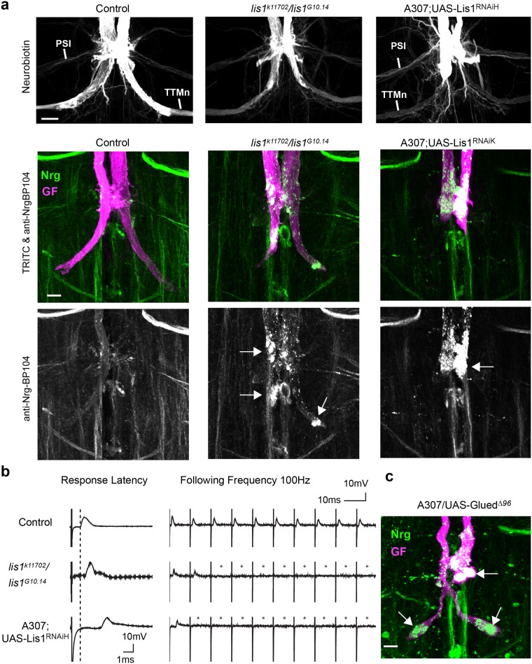
Fig 2. Lis1 phenotypes and Nrg localization in wild type and lis1 mutant backgrounds.
(a) Maximum intensity projection views of confocal image stacks showing representative GF terminals. GF synapse anatomy was visualized with neurobiotin (top panel) and TRITC-dextran (middle panel, magenta). Neuronal Nrg180 (green) was immunohistochemically labeled with monoclonal antibody BP104 (middle and lower panel). Neurobiotin dye-coupling of the GFs to the post-synaptic TTMns demonstrates the presence of synaptic connections between the medial TTMn dendrites and GF-TTMn. Accumulation of vesicular Nrg180 clusters in the synaptic terminal and in the axon at the PSI contact region are indicated with arrows. Scale bar is 10 μm. (b) Sample trace of electrophysiological recordings of the GF to TTM pathway from control animals and lis1 mutants. The response latency of wild type animals is approximately 0.8 ms (dashed line). Ten stimuli given at 100 Hz and the lack of some responses in mutant animals are indicated with asterisks. (c) Maximum intensity projection view of confocal image stacks of GF terminal expression a poisonous subunit of Drosophila dynactin p150 Glued protein. GF was dye injected with TRITC-dextran (magenta) and Nrg180 (green) was immunohistochemically labeled with monoclonal antibody BP104.

