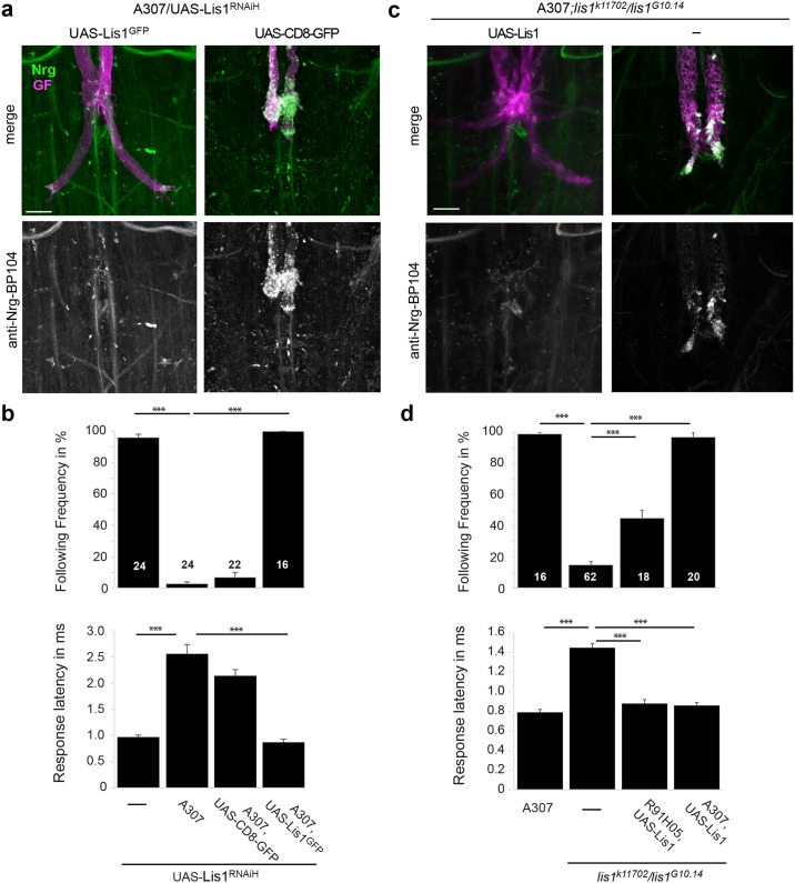
Fig 3. Rescue of Lis1 phenotypes.
(a, b) Rescue of Lis RNAi phenotypes with co-expression of Lis1GFP. Co-expression of UAS-CD8-GFP with UAS-Lis1RNAiH was used as a negative control. (c, d) Rescue of phenotypes in hypomorphic lis1k11702/lis1G10.14 with transgenic expression of wild type Lis1 in the GF and its target neurons or in the GF but not its target neurons with the A307 and R91H05 Gal4-drivers, respectively. A307, lis1G10.14/lis1k11702 without the UAS-Lis1 served as a negative control. (a, c) Giant fiber morphology and Nrg localization. All images show maximum intensity projections of confocal image stacks. GFs of adult flies were labeled by TRITC-Dextran injections (magenta) and displayed together with immunohistochemically labeled Nrg180 (anti-BP104 with goat anti-mouse-Cy5, pseudo-colored in green) in the VNC (upper row). Co-localization of both labels appears white. The lower row displays immunohistochemically labeled Nrg separately as gray scale images. Lis-GFP and CD8-GFP in (a) were present but are not shown. Scale bars are 20 μm. (b, d) The function of the GF synapse was determined with average following frequency and average response latency. Error bars indicate standard error of the mean and sample sizes are shown in the bars. Significant differences are indicated with asterisks (*** = p<0.001, Mann-Whitney Rank Sum Test).

