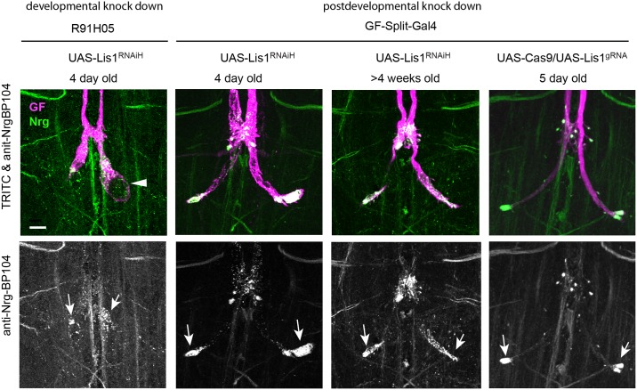
Fig 4. Phenotypes of GFs with post developmentally knocked down Lis1.
Comparison of phenotypes when Lis1 was knocked down in the GF during its development (R91H05) or after its development (GF-Split-Gal4) with RNAi (UAS-Lis1RNAiH) and CRISPR (UAS-Lis1gRNA). All images show maximum intensity projections of confocal image stacks. GFs of adult flies were labeled by TRITC-dextran injections (magenta) and displayed together with immunohistochemically labeled Nrg180 (green) in the VNC (upper rows). Co-localization of both labels appears white. The lower rows display immunohistochemically labeled Nrg180 separately as gray scale images. Scale bars are 20 μm. Localization of Nrg180 (anti-Nrg-BP104, green) in wild type and lis1 mutant backgrounds. Vesicular accumulations of Nrg180 in stunted and normal sized terminals are indicated by arrows. A large vacuole is indicated by an arrowhead.

