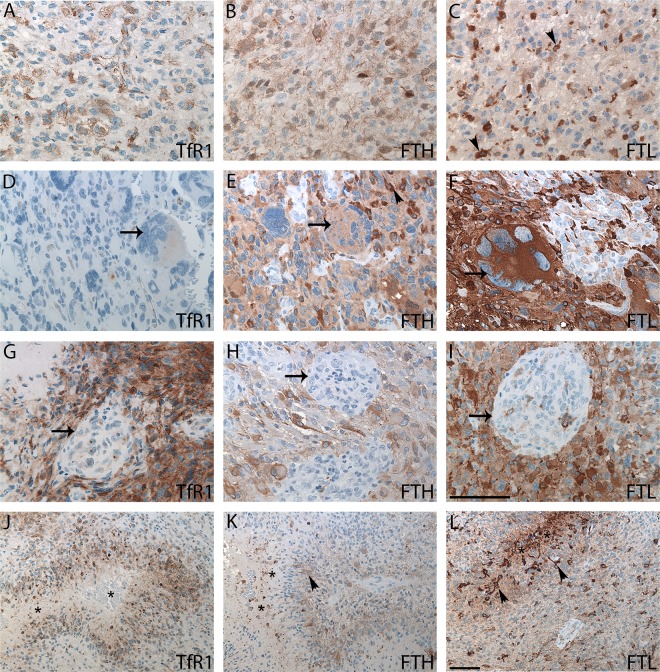
Fig 2. AAs and GBMs immunohistochemically stained with TfR1, FTH, and FTL.
AAs showed weak TfR1staining (A) and stronger FTH (B) and FTL (C) staining. GBMs with giant cells were weakly positive for TfR1 (D), moderately positive for FTH (E), and strongly positive for FTL (F). GBMs had no or limited glomeruloid staining for TfR1 (G), FTH (H), or FTL (I) (arrows). In GBMs with pseudo-palisading necroses (asterisks) TfR1-positive (J), FTH-positive (K), and FTL-positive (L) cells were observed in perinecrotic areas. Cells with microglial/macrophage morphology were easily identified in both the FTH (arrowheads) (E and K) and FTL stainings (arrowheads) (C and L). Abbreviations: AA anaplastic astrocytoma, FTH ferritin heavy chain, FTL ferritin light chain, GBM glioblastoma, TfR1 transferrin receptor-1. Scale bar: (A-I) 100 μm, (J-L) 100 μm.

