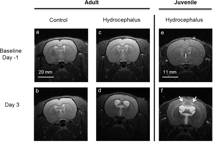
Fig 2. Typical axial high resolution T2-weighted anatomical MR images of adult and juvenile rats.
Control (a,b) and hydrocephalic adult rat (c,d) at baseline (Top row, one day prior to injection) and three days post-injection (bottom row). Kaolin injected into the cisterna magna enlarged the ventricles in the hydrocephalic rats. The size of the ventricles did not change in the sham injected controls. Fig 2 (e,f) show a typical MRI image of a juvenile rat at baseline and 3 days post injection. In the juvenile rats, the hyper-intense signal (see white arrows in f) was attributed to the presence of oedema in the white matter and inner layers of the cortical gray matter. In contrast, this was not observed in the adult hydrocephalic rats (see d). Brightness of all images was increased by 20% for visualization.

