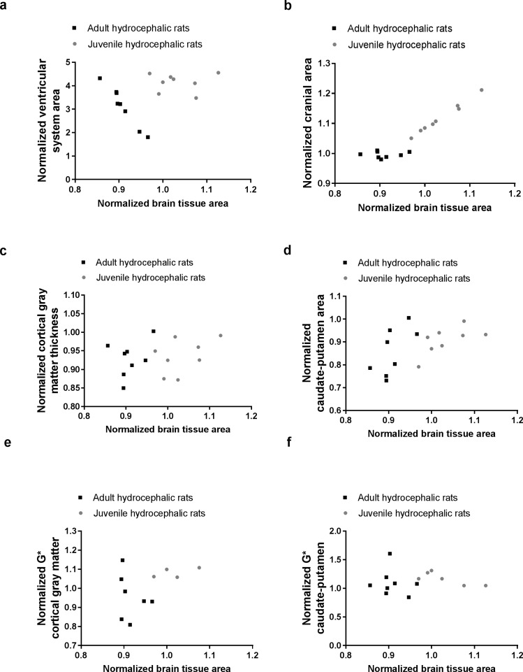
Fig 5. Effect of the brain compression on brain tissue deformation and stiffness in adult and juvenile hydrocephalic rats, three days post-hydrocephalus induction.
Relationships between the normalised brain tissue area and (a) normalised ventricular system cross-sectional area, (b) normalised cranial cross-sectional area, (c) normalised cortical gray matter thickness, (d) normalised caudate-putamen cross-sectional area, and normalised shear modulus (G*) of the (e) cortical gray matter and (f) caudate-putamen. Relationships were evaluated with Spearman correlations (Table 1). Note: In juvenile rats, four normalised G* measurements of the cortical gray matter and two of the caudate-putamen could not be calculated because the measurement was missing either at baseline or day 3.

