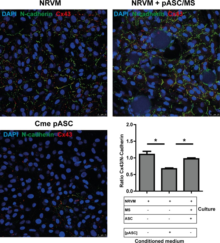
Fig 8. Immunofluorescence micrographs of cultures stained with N-cadherin and Cx43.
A: A monolayer of NRVM, a monolayer of NRVM cultured in pASC conditioned medium and a monolayer of NRVM co-cultured with pASC loaded microspheres are stained for N-Cadherin and Cx43. B:The ratio of Cx43: N-Cadherin in the various cultures, determined by the number of pixels. Ratios are based on 5–10 images taken in each of three independent cultures. * indicates p< 0.05. Abbreviations; Cme: conditioned medium, Cx43: connexin 43, MS: microspheres, NRVM: neonatal rat ventricular myocytes and pASC: porcine adipose tissue-derived stromal cells.

