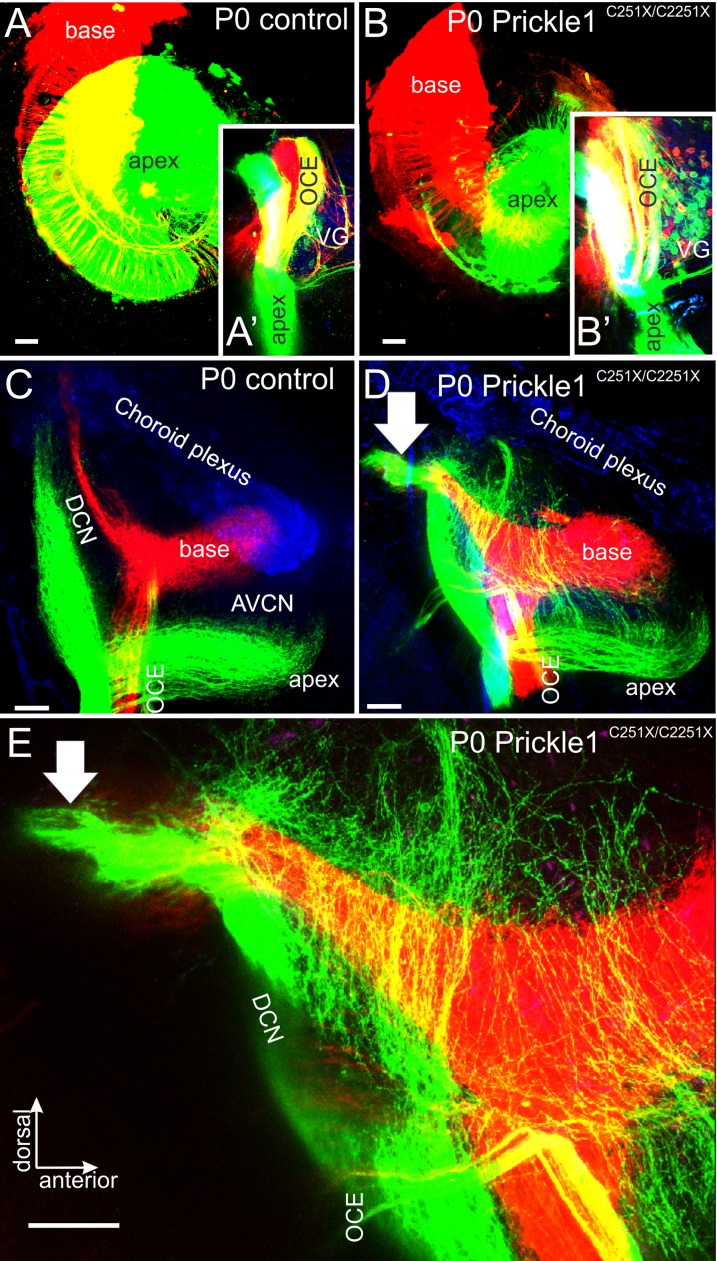
Fig 4. Central projections from apical afferents are expanded in the cochlear nuclei in Prickle1C251X/C251X mutants.
Different colors of lipophilic dyes were applied to apex and the base of the cochlear (A, B), and their central projection were analyzed (A’, B’, C-E). (A and B) Overview of the cochlea showing the application of red dye to the base and green dye to the apex in control (A) and Prickle1C251X/C251X mutant (B). After 3 days of diffusion, there was partial overlap of the dye. (A’ and B’) Selective bundles of afferents and olivocochlear efferents (OCE) passed along the vestibular ganglion (VG). Only in Prickle1C251X/C251X mutant (B’), OCE separated into several bundles. In addition, several vestibular ganglion neurons (VG) were labeled. (C-E) Projections to the cochlear nucleus of the control (C) and the Prickle1C251X/C251X mutant (D, E). (C) In controls, afferent bundles from the apex and the base segregated and formed distinct fascicles. (D, E) In the mutant, although afferents from the base projected normally to the dorsal cochlear nucleus complex DCN, afferents from the apex formed collaterals that spread out throughout the DCN and the anterior-ventral cochlear nuclei (AVCN). (E) Higher magnification of the DCN of (D), showing details of apical afferents passing basal turn afferents to branch in the most dorsal aspect of the cochlear nucleus complex. Arrow, afferents innervating vestibular nuclei. Scale bars, 100 μm.

