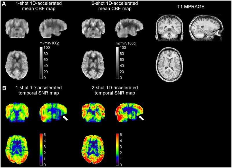
Fig 5. (A) Mean CBF and corresponding (B) temporal SNR maps obtained with 1-shot and 2-shot 1D-accelerated readouts in Study 2 from a representative subject, alongside the anatomical dataset.
Note how the two-shot readout resulted in both an increase in SNR and a further reduction in through-plane blurring, evident in the coronal and sagittal views. White arrows point at orbitofrontal areas, which typically suffer from low SNR due to shortened T2* and T2. CBF units are in ml/min/100g.

