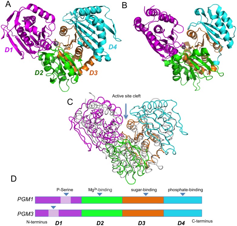
Fig 2. Crystal structures of human PGM1 and a PGM3 homolog.
(A) Human PGM1 (PDB ID 5EPC) colored by structural domain: domain 1 (magenta, residues 1–191); domain 2 (green, residues 192–304); domain 3 (orange, residues 305–421), and domain 4 (cyan, residues 422–562). Bound metal ion in the active site is shown as gray sphere. (B) A homolog of PGM3 (N-acetylphosphoglucosamine mutase from C. albicans; PDB ID 2DKD) colored as in (A). The domains are: domain 1 (residues 1–191); domain 2 (residues 192–311); domain 3 (residues 312–456); and domain 4 (residues 457–544). (C) A structural superposition of PGM1 (colored by domain) and the PGM3 homolog (white). (D) A schematic of the domain arrangement of PGM1 and PGM3, indicating the residues transposed by the circular permutation in domain 1 of PGM3. Key active site regions are indicated.

