
Fig 3. Sequence preferences for key functional regions of the PGM1 paralogs.
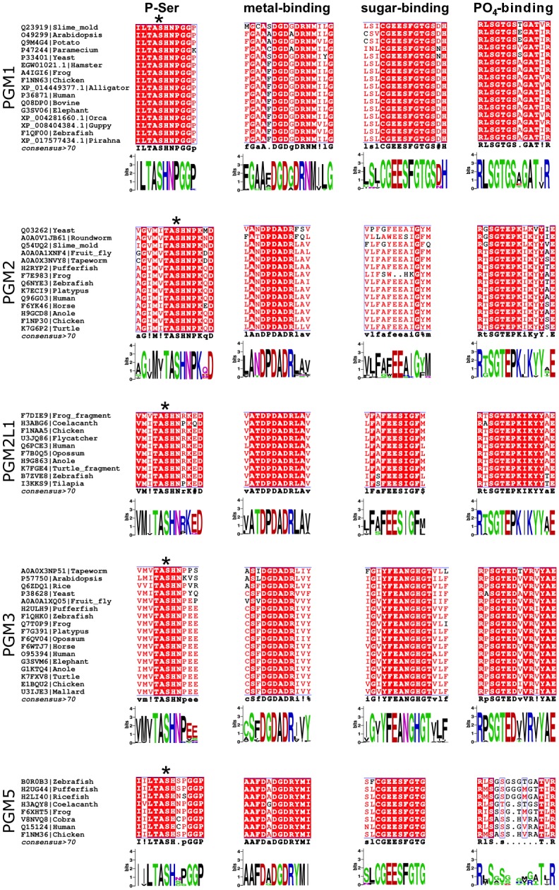
Selected regions of paralog-specific multiple sequence alignments are shown for the phosphoserine (P-Ser), metal-binding, sugar-binding, and PO4-binding loops. Identical residues are highlighted with red background; similar residues are shown in red font. Asterisk (*) indicates the catalytic serine residue.