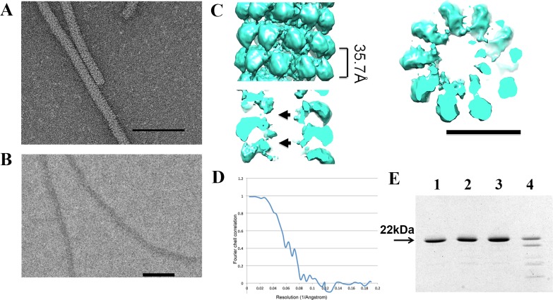
Fig 2. EM analysis of AltMV virus-like particles (VLPs).
А. TEM of AltMV VLPs. Samples were stained with 2% uranyl acetate. Bar– 100 nm. B. Cryo-electron microscopy of AltMV VLPs. C. 3D reconstruction: on the left–surface of AltMV VLP, on the right–horizontal slice of AltMV VLP, below–vertical slice of AltMV VLP. Arrows are pointing to the absence of the density, associated to the viral RNA in Fig 1. D. FSC curve. E. Structure analysis by trypsin treatment. 1 –AltMV virions, 2 –AltMV virions treated by trypsin, 3 –AltMV VLPs, 4 –AltMV VLPs treated by trypsin. All samples contained 2 μg of material. Analysis in 8–20% SDS-PAGE, gel was stained with Coomassie G-250.

