
Fig.11.
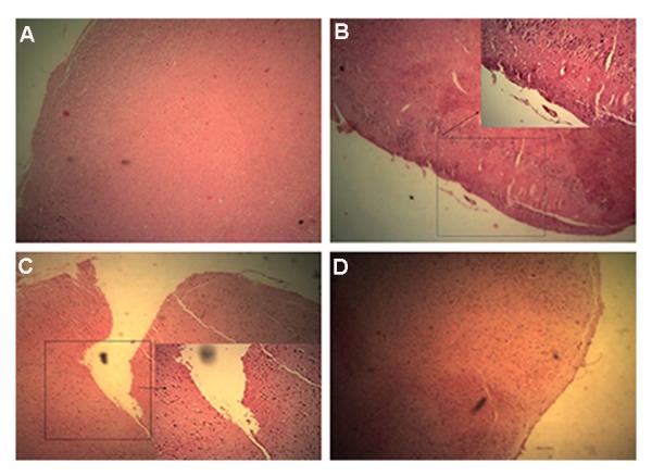
Immunohistology analysis. H&E staining of brain in: A. Normal mice without brain lesions and cell infiltration foci, B. Experimental autoimmune encephalomyelitis (EAE) induced mice with no treatment Fcondensed cell infiltration foci and brain lesion are visible, C. EAE induced mice were treated with WJSCs again cell infiltration and brain lesions are visible but less that untreated group, and D. EAE induced mice treated with WJSC transducted with transfer vector shows more reduction of brain lesions and cell infiltration foci.