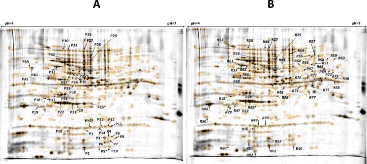
Fig 1.
Fused images of three Pro-Q Diamond (a) and three RuBPS (b) stained gels. The arrows point to spots with significantly altered normalized spot volumes in the cappz1 mutant C. albicans strain. Letters P or R in front of spot IDs indicate the staining methods. In the spots labelled with * more than one protein was found.

