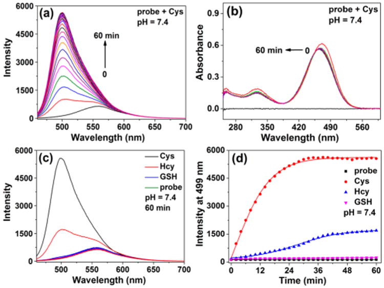
Figure 1.

(a) Time-dependent fluorescence emission spectra of 1 (30 μM) in the presence of 10 equiv of Cys in Hepes buffer/DMSO (1:1, v/v, pH 7.4) at 25 °C. (b) Corresponding time-dependent UV–vis spectra of 1 (30 μM) in the presence of 10 equiv of Cys in Hepes buffer/DMSO (1:1, v/v, pH 7.4) at 25 °C. (c) Fluorescence emission spectra of 1 (30 μM) upon addition of 10 equiv of Cys, Hcy, GSH, Ala, Asn, Arg, Asp, Gln, Glu, Gly, His, Ile, Leu, Lys, Met, Phe, Pro, Ser, Thr, Trp, Tyr, and Val in Hepes buffer/DMSO (1:1, v/v, pH 7.4) at 25 °C. (d) Time-dependent fluorescence emission intensity changes of 1 toward 10 equiv of biothiols at 499 nm (λex = 447 nm, slit: 5 nm/5 nm).
