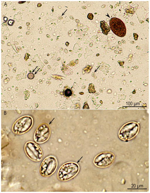
Figure 1.
Sporocysts of Sarcocystis spp. from a bobcat feces, processed following sugar flotation, Unstained. (A) Lower magnification. Note a trematode (Paragonimus sp.) ova (arrowhead), coccidian oocyst (double arrows), and many Sarcocystis sporocysts (arrows) that lie at different focus than the trematode ova. (B) Higher magnification to show Sarcocystis sporocysts (arrows). Sporocysts of many species of Sarcocystis overlap in dimensions (8 × 15 μm).

