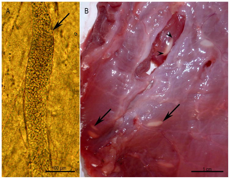
Figure 3.
Microscopic and macroscopic sarcocysts, Unstained. (A) Microscopic sarcocyst (arrow) in the skeletal muscle of a naturally infected bobcat. (B) Macroscopic sarcocysts (arrows) on the surface of the esophagus of naturally infected water buffalo. Note the cysts are covered with fascia (arrow) and one of them is exposed (arrowheads) (Courtesy of Dr. M. A. Hilali, Cairo University, Egypt).

