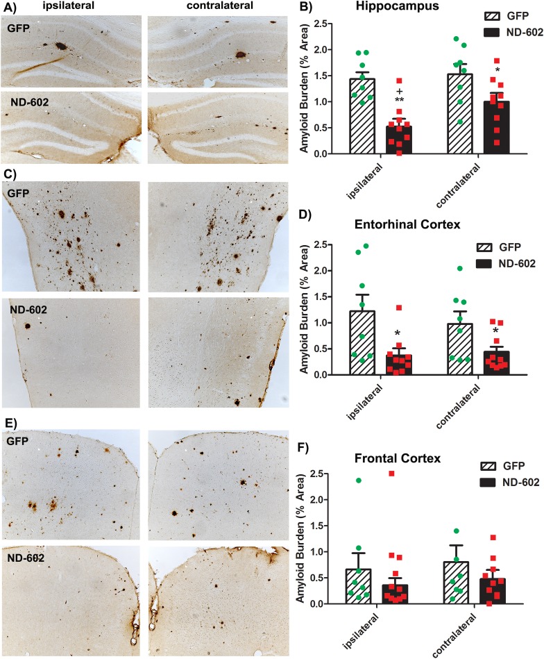
Fig 3. Beta-amyloid burden following ND-602.
(A,C,E) Representative photomicrographs depicting β-amyloid immunolabeling in the (A) hippocampus, (C) entorhinal cortex, and (E) frontal cortex of a Tg2576 mouse brain following unilateral intracerebral administration of either LV-GFP, or LV-PGRN (ND-602). Amyloid burden was significantly reduced in the (B) dentate gyrus and (D) entorhinal cortex of those animals receiving ND-602 administration. (F) Apparent reductions in amyloid burden observed in the frontal cortex failed to reach statistical significance due to a high degree of variability in deposition in this region, at this time point. Each bar represents the mean (± S.E.M.) (n = 8–10) amyloid burden (% area) measured across 4 coronal sections. ** sig. diff. from GFP-treated controls, p < 0.001; * p < 0.05 + sig. diff. from contralateral hemisphere, p < 0.05.

