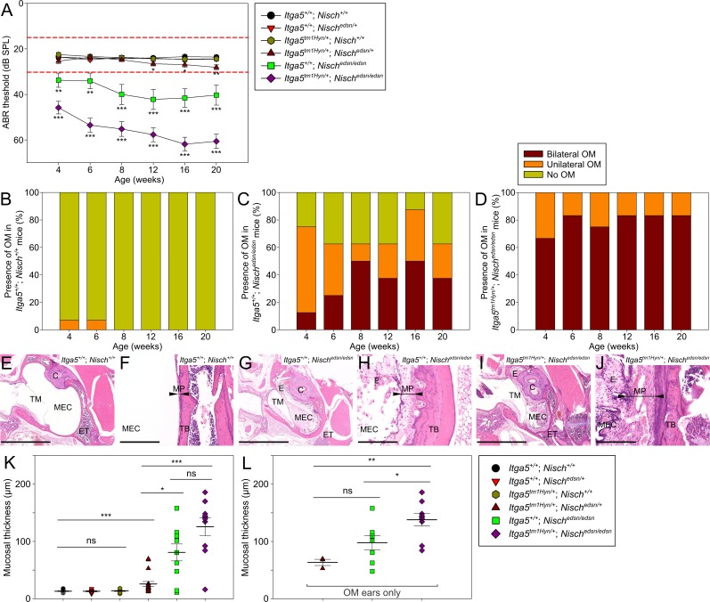
Fig 5. Deficiencies in Nisch and Itga5 exacerbate the otitis media phenotype.
(A) Click-evoked ABR thresholds across a time course show Itga5tm1Hyn/+; Nischedsn/edsn mice exhibit significantly elevated auditory thresholds compared to Itga5+/+; Nischedsn/edsn mice. Additionally, a mild late-onset hearing deficit is observed in Itga5tm1Hyn/+; Nischedsn/+ mice, with onset at 12 wk. Expected ABR threshold range for normal hearing was between 15–30 dB SPL (dashed red lines). Itga5+/+; Nisch+/+ n = 14; Itga5+/+; Nischedsn/+ n = 14; Itga5tm1Hyn/+; Nisch+/+ n = 15; Itga5tm1Hyn/+; Nischedsn/+ n = 13; Itga5+/+; Nischedsn/edsn n = 8; Itga5tm1Hyn/+; Nischedsn/edsn n = 12. (B-D) Visual inspection of the tympanic membrane was used as a semi-quantitative measure for the prevalence of OM. (B) Itga5+/+; Nisch+/+ mice show a very low prevalence of unilateral OM at 4 wk and 6 wk only. (C) Itga5+/+; Nischedsn/edsn mice show a progressive increase in prevalence of OM, whereas (D) Itga5tm1Hyn/+; Nischedsn/edsn mice display a consistently high prevalence of bilateral OM throughout the time course. (E-J) H&E stained transverse sections of the MEC and mucoperiosteum, in 20 wk (E, F) Itga5+/+ Nisch+/+, (G, H) Itga5+/+; Nischedsn/edsn and (I, J) Itga5tm1Hyn/+; Nischedsn/edsn mice. Both Itga5+/+; Nischedsn/edsn and Itga5tm1Hyn/+; Nischedsn/edsn mice demonstrate chronic inflammation with an exudate. Inflammation of mucosa was more severe in sections from Itga5tm1Hyn/+; Nischedsn/edsn ears, with increased polypoid exophytic growths and a thick cellular effusion. (K) Blinded assessment of mean mucosal thickness demonstrates significant increases in Itga5tm1Hyn/+; Nischedsn/+, Itga5+/+; Nischedsn/edsn and Itga5tm1Hyn/+; Nischedsn/edsn mice compared to wild-type. Both Itga5+/+; Nischedsn/edsn and Itga5tm1Hyn/+; Nischedsn/edsn mice exhibit significant increases in mucosal thickness compared to Itga5tm1Hyn/+; Nischedsn/+ mice. Itga5+/+; Nisch+/+ n = 12; Itga5+/+; Nischedsn/+ n = 8; Itga5tm1Hyn/+; Nisch+/+ n = 10; Itga5tm1Hyn/+; Nischedsn/+ n = 18; Itga5+/+; Nischedsn/edsn n = 10; Itga5tm1Hyn/+; Nischedsn/edsn n = 10. (L) To account for the disparities in OM prevalence, the mean mucosal thickness was additionally assessed for OM ears only. Itga5tm1Hyn/+; Nischedsn/edsn OM ears exhibit significant increases in mucosal thickness compared to Itga5+/+; Nischedsn/edsn OM ears. OM only: Itga5tm1Hyn/+; Nischedsn/+ n = 3; Itga5+/+; Nischedsn/edsn n = 8; Itga5tm1Hyn/+; Nischedsn/edsn n = 9. C, cochlea; ET, eustachian tube; E, exudate; MEC, middle ear cavity; MP, mucoperiosteum (arrowheads); TB, temporal bone; TM, tympanic membrane. E, G, I scale bar = 2 mm; F, H, J scale bar = 200 μm. ns P > 0.05; * P < 0.05; ** P < 0.01; *** P < 0.001. Error bars indicate standard error of mean. A Kruskall-Wallis test was performed followed by Dunn’s multiple comparison tests for post-hoc analysis.

