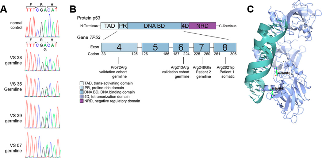
Figure 3. Germline TP53 variants.

A. Sanger sequencing of exons 2–11 of TP53 was performed on the germline DNA of 37 pediatric cancer survivors who subsequently developed radiation-induced SMNs. Shown are chromatograms from the four individuals demonstrating the germline variant c.A639G (p.R213R), which is a synonymous single nucleotide polymorphism rs1800372. The trinucleotide codon is shown in color below the single letter amino acid symbol. B. Schematic of the p53 protein, indicating the locations of germline and somatic variants associated with SMNs. C. Three-dimensional structure of the p53 protein indicating the positions of the SMN-associated mutated amino acids.
