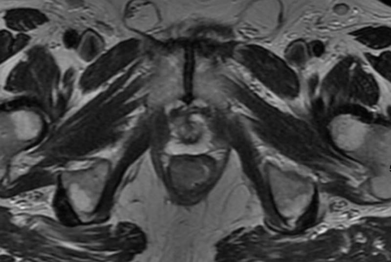
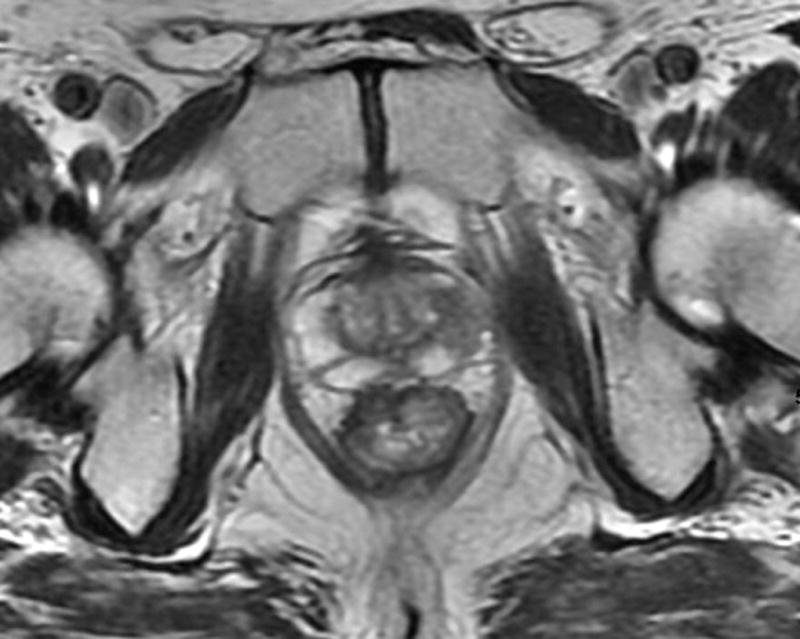
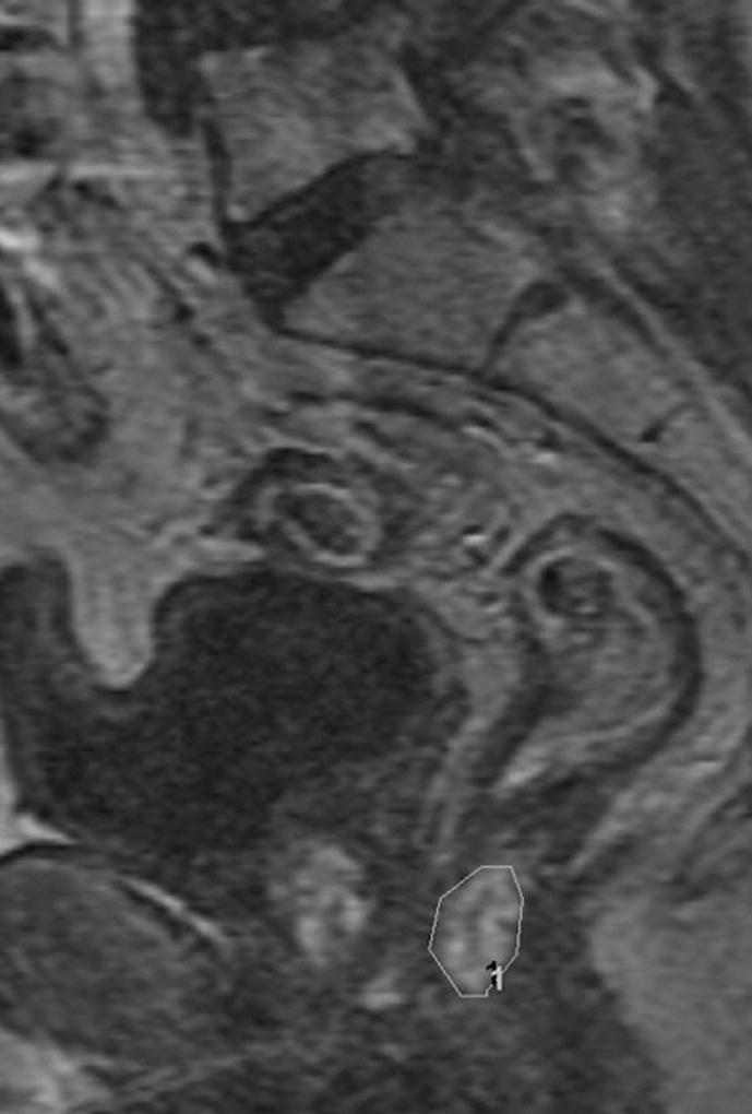
Fig. 2. 66-year-old man with clinical T3N0, low rectal cancer.


a. T2-weighted axial baseline MRI through lower rectum showing mass filling rectal lumen posterior to prostate
b. T2-weighted axial post-chemoradiotherapy MRI through lower rectum showing scar and questionable residual tumor at right anterior wall in former position of tumor
c. Sagittal DCE-MRI post chemoradiotherapy with region of interest on early enhancing tissue in tumor bed with Ktrans90 = 0.35 and 55% tumor response on subsequent histopathology
