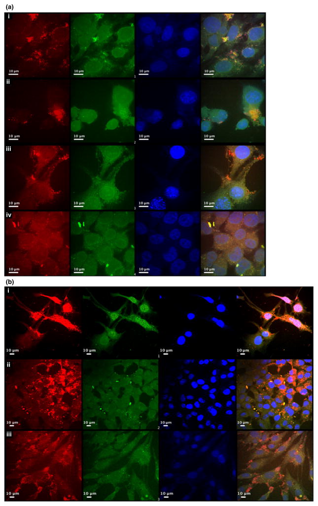
Fig. 3.
Confocal microscopy evidence for delivery of siRNA conjugated JB577 QD into cytosol. (a) Cells were transfected with polyethylene glycol (PEG) QD-JB577-siRNA constructs for 24 h, siRNA was labeled with Cy3 (green), quantum dots are shown as red and nuclei stained with 4′,6-diamidino-2-phenylindole (DAPI) (blue). The cells were then confocal imaged with a 100× oil immersion lens. The cell types were as follows (i) 102R fibroblasts (ii) human oligodendroglioma (iii) human cerebral epithelial cells (HCEC) (iv) HCT-116. (b) Cells were transfected with PEG QD-JB577-siRNA constructs for 24 h, siRNA was labeled with Cy3 (green), quantum dots are shown as red and nuclei stained with DAPI (blue). The cells were then imaged with a 40× oil immersion lens. The cell types were as follows (i) 102R (ii) HCT-116 (iii) HCEC.

