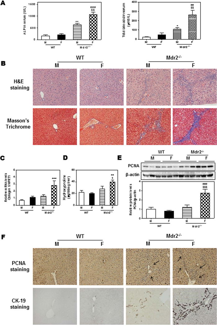
Figure 1. Gender disparity of cholestatic liver injury in Mdr2−/− mice.

100-day-old Mdr2−/− mice and age-matched FVB WT mice were sacrificed. (A) Serum levels of ALP and total bile acids (TBA). (B) Representative images of H&E and Masson’s Trichrome staining. (C) The mRNA level of collagen I was determined by real-time RT-PCR and normalized using HPRT1 as an internal control. (D) Hepatic hydroxyproline content. (E) Protein level of PCNA was determined by Western blot analysis and normalized with β-actin as an internal control. Representative images are shown. (F) Representative images of immunohistochemistry staining of PCNA and CK-19. Statistical significance: *P< 0.05; **P<0.01, compared with male (M) WT (WT) mice; ##P<0.01; ###P<0.001, compared with female (F) WT mice; $P < 0.05, $$P<0.01; $$$P<0.001, compared with (M) Mdr2−/− mice (n=6).
