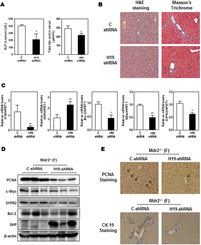
Figure 7. Down-regulation of H19 reduced cholestatic injury in female Mdr2−/− mice.

Female (F) Mdr2−/− mice at age 60-day were injected with purified control adenovirus or H19 shRNA adenovirus (2 ×10¹⁰ virus particles per mouse). Mice were harvested after one week. (A) Serum ALP and TBA levels. (B) Representative images of H&E and Masson’s Trichrome staining. (C) The relative mRNA levels of H19, SHP, Bcl-2, ERα and S1PR2 were determined by real-time RT-PCR and normalized using HPRT1. (D) Representative images of Western blot analysis of PCNA, c-Myc, S1PR2, Bcl-2 and SHP normalized with β-actin as an internal control. (E) Representative images of immunohistochemistry staining of PCNA and CK-19 in livers. Statistical significance: *P<0.05, **P<0.01, compared with control shRNA group (n=6).
