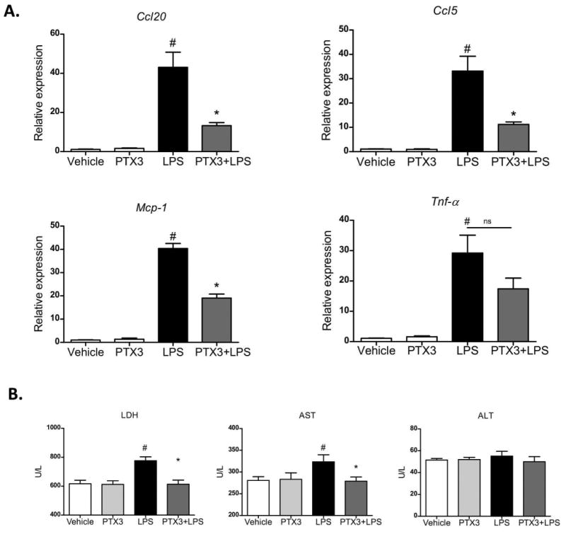
Figure 4. Effect of PTX3 in high precision-cut liver slices.

Liver slices from mice treated with CCl4 were incubated with or without PTX3 and stimulated with LPS. a) Gene expression of Ccl20, Ccl5, Mcp-1 and Tnf-α in liver slices after incubation for 1 hour with rPTX3 (500 ng/mL) (n=4) or vehicle with or without addition of LPS (25 μg/mL) (n=4) for an additional 6 hours. #p<0,05 compared with control; *p<0,05 compared with LPS induction. b) Tissue culture supernatant levels of lactate dehydrogenase (LDH), aspartate aminotransferase (AST), and alanine aminotransferase (ALT) in (n=4) from cultures of liver slices pre-incubated with rPTX3 or vehicle for 1 hour before LPS stimulation for 6 hours #p<0,05 compared with control; *p<0,05 compare with liver slices treated with LPS.
